What imaging does a pituitary surgical team actually need to see?
Pituitary surgery is not planned from a written report alone. The operating surgeon needs the actual image files, not only the radiologist's summary, because the decision depends on where the tumour sits relative to the optic chiasm, the cavernous sinuses, the sphenoid sinus and the normal pituitary gland. A report may describe a macroadenoma as 'suprasellar', but the surgeon still needs to see how far it extends, whether it compresses the optic apparatus, and what route through the nose is realistic.
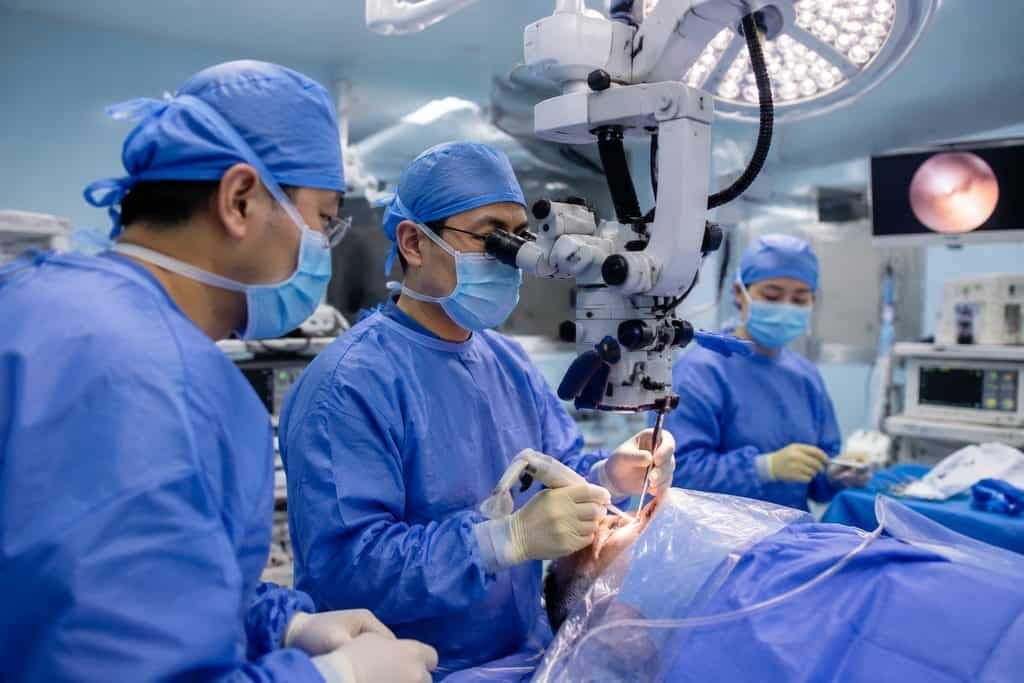
Endoscopic transsphenoidal pituitary surgery reaches the pituitary region through the nose. That approach makes the nasal and sinus anatomy part of the surgical plan, so the team will want to review the sinus anatomy on the same imaging set. If your images were done elsewhere, ask whether the hospital can accept the original DICOM files on disc or through a secure upload, and whether it needs the images repeated in China. Do not assume a repeat is required; do not assume your existing images are sufficient either. That is a question for the receiving surgical team.
The practical point is that imaging is not a single test result. It is a dataset that a surgeon reads alongside your hormone results, your visual field testing and your symptoms. A missing sequence, a low-resolution study or a report without images can slow the review even when the diagnosis itself is already clear.
MRI, CT and visual field testing: different questions, different records
MRI is the main imaging method for defining a pituitary tumour and its relationship to soft tissue structures. CT may be requested for bony anatomy, particularly the sphenoid sinus and skull base, when the surgical route is being planned. These are not competing tests; they answer different questions. If you have had both, keep both. If you have had only one, ask the surgical team whether the other is needed for your case rather than arranging it yourself.
Visual field testing is not imaging, but it is part of the same assessment. A tumour that presses on the optic chiasm can change peripheral vision before the patient notices it. The surgical team may want formal visual field results, not just a comment that vision seems fine. If you have had visual field testing, include the actual charts or numeric results. If you have not, ask whether it should be arranged locally before travel or whether the hospital will arrange it during assessment.
Keep the original imaging files and the written reports together. Reports in a language the receiving team cannot read are less useful than the images themselves, but a translated summary can help the coordinator route your file to the right specialist. Do not send a complete medical archive in your first message; a short summary with the key reports is enough to start.
Why endocrine results belong in the same review as the images
A pituitary tumour is not only a structural problem. Some tumours produce excess hormones, some reduce normal pituitary function, and some do neither. The imaging shows where the tumour is; the endocrine results show what it is doing. A surgical team deciding on an operation needs both, because a hormone-producing tumour may need medical treatment before or alongside surgery, and a patient with low cortisol or thyroid hormone may need that addressed before any operation is considered.
This is why the question 'is my MRI enough?' has no complete answer on its own. The same image can lead to different plans depending on the hormone profile. Bring your endocrine blood results with dates and units, any dynamic testing results, and any medication list. If you have not had endocrine testing, ask the receiving clinician whether it should be done locally first or whether the hospital will arrange it.
Hormone and vision issues can be relevant to assessment. That does not mean every patient needs the same tests, and it does not mean you should start, stop or change any medication on your own. Steroid and hormone replacement decisions belong to the treating clinician, who needs to see your results and your current prescriptions.
Questions to put to the surgical team about your images
The most useful thing you can do before an overseas consultation is to ask precise questions and write down the answers. Vague questions produce vague replies. Instead of asking 'do you need my scans?', ask which sequences and which time period they want, whether they will accept images from your hospital, and whether a repeat study would be arranged in China if the existing images are not adequate.
It also helps to ask who reads the images. In a complex pituitary case, the review may involve a neurosurgeon, an endocrinologist and sometimes an ophthalmologist or ENT surgeon. Ask how those assessments are coordinated, whether they happen on the same visit or across several appointments, and who takes responsibility for the final surgical recommendation. You are not asking the coordinator to make a clinical decision; you are asking how the hospital organises one.
Finally, ask what happens after surgery. Who will manage ongoing hormone follow-up, and who will arrange surveillance imaging? Pituitary care often continues for years, and the answer may involve your local clinicians as well as the team in China. Ask for the plan in writing so your doctors at home can continue it.
- Which MRI sequences and which date range does the surgical team want?
- Will the hospital accept DICOM files from my hospital, or would imaging be repeated in China?
- Is CT of the sinuses or skull base needed for my case, and who decides?
- Are visual field results required before the surgical review?
- Which specialties will review my case, and how is that coordinated?
- Who will manage hormone follow-up and future imaging after I return home?
What imaging cannot tell you, and what to confirm instead
Imaging can show the size and position of a pituitary tumour. It cannot by itself tell you whether surgery is the right choice, when it should happen, or what your recovery will be. Those judgements depend on the tumour type, hormone function, your symptoms, your vision and your general health. A scan that looks impressive may need no operation; a smaller tumour causing hormone excess may need treatment. The images inform the decision; they do not make it.
This is also why no article can tell you whether your images are 'good enough' for surgery in China. The receiving hospital decides whether to accept your case, what further assessment it needs, and what it will recommend. A records-based opinion can help you understand the options, but it does not establish hospital acceptance or a final treatment plan. If your vision is worsening or you have sudden severe headache, visual loss or confusion, seek urgent local care rather than waiting for an overseas reply.
On the practical side, do not assume that a consultation, a scan and a surgical decision can all be compressed into one trip. Ask the hospital how it sequences assessment and what it expects from you at each stage. If you need a written estimate, ask what the quote includes and what remains undecided; the hospital, not a coordinator, provides that scope.
A practical next step for your imaging question
Start by gathering the essentials: your most recent pituitary MRI images and reports, any CT of the sinuses or skull base, your visual field results if available, and your endocrine blood results with dates and units. Write a short summary of your main question, for example whether your existing imaging is sufficient for a surgical review or whether further assessment would be arranged in China.
You can send that summary through the enquiry form, email or WhatsApp. An initial enquiry is free and does not require buying a proxy consultation. Our team checks the available diagnosis, records and your main question, identifies missing information and suggests the relevant next step. That is not a diagnosis and not a promise of acceptance. The hospital and its clinicians decide suitability, further imaging and treatment.
If you want background on the procedure itself, the pituitary tumor surgery reference page explains the broader assessment and treatment context. Use it alongside this guide, not instead of the specific answers from the treating team about your own images.
Sources & scope of this guide
References and official service information relevant to this guide.
This is general planning information and has not been individually reviewed by a doctor. Medical decisions and personal treatment advice come from your treating clinicians.