
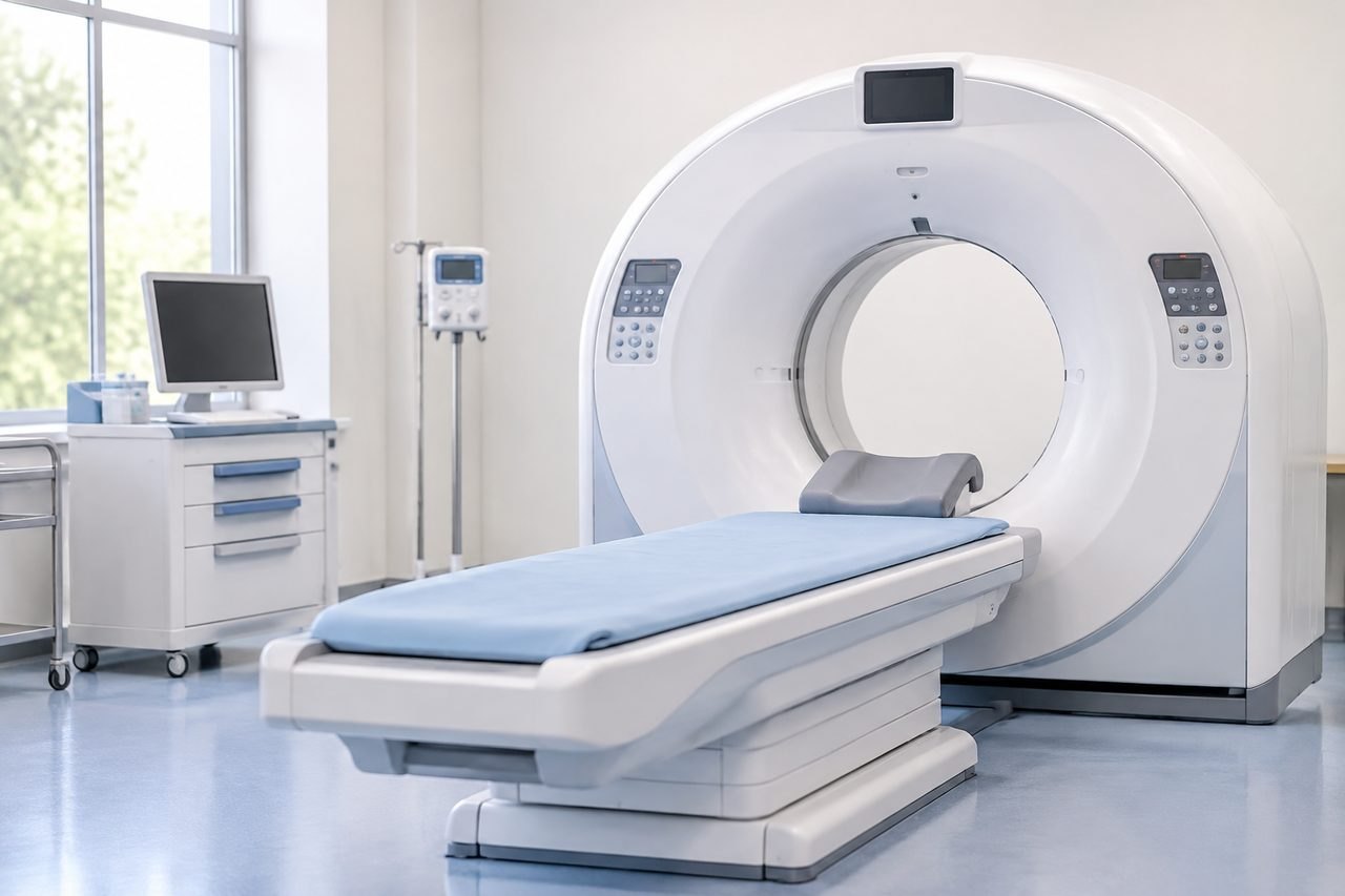
Why imaging is a secondary question in most leukemia reviews
Leukemia is a disease of blood and bone marrow, so the core evidence in a review is usually laboratory and pathology material rather than pictures. A hematology team reviewing a case will typically want to see the diagnostic blood counts, the bone marrow aspirate and biopsy report, flow cytometry, cytogenetics and molecular testing, plus the treatment history and current status. Imaging supports that picture; it does not replace it.
That matters for planning. A patient who arrives with a folder of scans but no marrow report, no cytogenetic result and no treatment summary has brought the least useful part of the record. A patient who sends the laboratory and pathology documents first gives the reviewing team something to reason from, and the imaging question can then be answered specifically rather than guessed at.
The type of leukemia changes the imaging question. In acute leukemia, imaging is often used to look for specific complications or to assess organs before intensive treatment, not to diagnose the disease itself. In chronic leukemias the review may hinge on disease phase, prior therapies and molecular response. The same scan request does not apply across these situations, and no article can substitute for the treating team's assessment of an individual case.
Which imaging questions a hematology team may actually ask
Rather than asking "what scans do I need", it is more useful to understand the categories of imaging question that come up in leukemia care. Each one exists to answer something specific, and a scan done without a clear question adds cost and delay without adding information.
One common category is assessment before or during intensive treatment. A treating team may want to know about organ function, prior lung disease or an existing infection risk before certain therapies. Another category is investigation of a specific symptom: persistent fever, chest symptoms, bone pain, neurological signs or an unexplained finding on examination. In those situations imaging is driven by the clinical problem, not by the leukemia label.
A third category is staging-type assessment in conditions where the team needs to know whether disease is present outside the blood and marrow. This is more relevant in some leukemia subtypes and in lymphoma than in others, and it is a decision the hematologist makes with the specific diagnosis in hand. A fourth category is monitoring a known complication over time, such as a lung finding or an infection being treated.
The practical consequence is that you should not commission scans in advance to "complete" a file. Ask the receiving team, in writing if possible, which imaging, if any, they want for the question your case raises, and whether they will accept imaging done at an accredited facility near home. Whether a Chinese hospital will accept outside imaging, and under what conditions, is something to confirm with that hospital rather than assume.
The records that make an imaging decision possible
A reviewing hematologist cannot say whether imaging is needed without the underlying diagnosis and treatment context. The most useful first package is therefore documentary, not radiological. It should let a clinician understand what type of leukemia this is, how it was confirmed, what treatment has been given, how the disease has responded, and what the current clinical question is.
Pathology and laboratory material usually sits at the centre of that package. The original bone marrow report, flow cytometry, cytogenetics and any molecular results are the documents that define the disease. If a review is being sought because the diagnosis or subtype is uncertain, a pathology re-review may be more valuable than any new scan, and the relevant reference explains how that process works.
Treatment records matter just as much. A chronological summary of regimens, dates, responses, complications and current medicines lets the team see the trajectory. So does a clear statement of the patient's own question: is this about confirming a diagnosis, considering a different treatment, seeking a transplant opinion, or understanding a complication? The answer changes what the team will ask for next.
Imaging that has already been done should be included, but in a usable form. Reports alone are often less informative than the images themselves, and whether a hospital can load outside images from discs or a portal is a question for that hospital. Do not send a complete archive in a first message; a brief summary and a list of what exists is enough to start, and the team can then request specific items.
What imaging cannot settle, and where the limits lie
It is worth being clear about the boundaries. Imaging does not establish a leukemia diagnosis, does not measure molecular response, and does not replace marrow examination when the question is about disease in the marrow. A normal scan does not exclude active leukemia, and an abnormal scan does not confirm it. Those judgements rest on laboratory and pathology evidence interpreted by the treating hematologist.
There is also a safety dimension. Contrast studies, sedation and radiation exposure each carry their own considerations, and whether a particular scan is appropriate depends on kidney function, allergies, pregnancy status and the clinical question. These are decisions for the treating clinicians, who can weigh whether the expected information justifies the test. A coordination service cannot make that judgement, and should not be asked to.
Finally, a records-based review has limits that patients should understand before paying for anything. A specialist reviewing documents from a distance can comment on the material available and identify what is missing, but cannot examine the patient, cannot order tests in China on the patient's behalf, and cannot promise that a hospital will accept the case or that a particular treatment will be available. Eligibility for a clinical trial, a transplant pathway or a specific therapy is established only through the relevant formal assessment, not through an initial review.
Planning the practical side without inventing a timetable
If the review leads toward care in China, the imaging question becomes part of a wider practical plan. Ask the hospital, through whatever channel you use, which investigations it would repeat or require locally, whether it accepts outside results, and how those decisions are documented. Answers vary by hospital and by case, so a written reply about your own situation is more useful than general expectations.
Language and record handling are worth settling early. If reports and images are in another language, ask whether interpretation is needed and who provides it. If you use a coordination service, be clear about what it does and does not do: it can help with appointment requests, hospital navigation and interpretation, but clinical decisions, test ordering and acceptance remain with the hospital and its clinicians. Coordination fees and hospital charges are separate, and you should ask for the written scope of any quote rather than rely on an estimate.
For patients who want a specialist's view before travelling, a records-based opinion is one route, and it is optional. It is not a prerequisite for every appointment, and an initial enquiry does not require buying one. The free initial review checks what diagnosis and records are available and what the main question is; it is not a diagnosis and does not promise acceptance.
A sensible next step
Start with the documents that define the disease rather than with scans. Gather the diagnostic marrow and pathology reports, cytogenetics and molecular results, a treatment chronology and a one-paragraph statement of your question, then send a short summary through the enquiry form, email or WhatsApp. The team can tell you what is missing and whether a specialist review or an appointment request is the sensible next step. Keep any imaging reports you already have in reserve for when a clinician asks for them specifically.
If you want to understand the wider treatment context first, the acute leukemia treatment reference and the hematology specialty page set out how care for blood cancers is organised. Neither replaces an individual assessment, and neither establishes that any particular therapy is available for your case.
Sources & scope of this guide
References and official service information relevant to this guide.
- hematology
- Related treatment reference
- NCI: Adult Acute Myeloid Leukemia Treatment (PDQ) – diagnosis and staging evaluation
- NCI: Chronic Lymphocytic Leukemia Treatment (PDQ)
- NICE guideline NG12: Suspected cancer – recognition and referral
- American College of Radiology: Appropriateness Criteria
This is general planning information and has not been individually reviewed by a doctor. Medical decisions and personal treatment advice come from your treating clinicians.