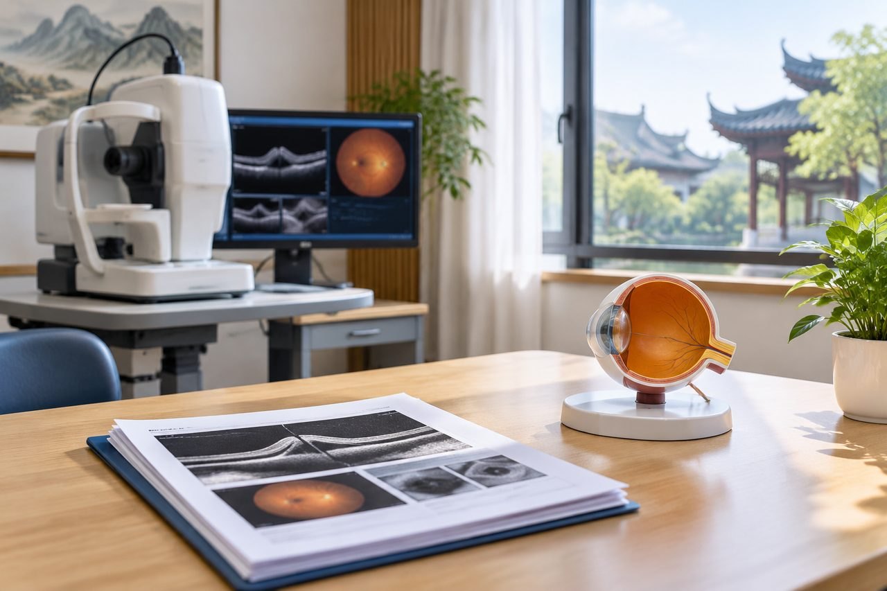
Why the OCT report matters more than the diagnosis label
A diagnosis of macular hole tells you that the central retina is affected. It does not tell a surgeon what the hole looks like now, how long it has been present, or whether the surrounding retina is stable. The OCT scan is the record that carries those distinctions. When you send records to a hospital in China, the treating team reads the images and measurements, not just the words on the report.
This is why two patients with the same diagnosis label can receive different advice. A small hole with attached edges and a large hole with lifted edges are different surgical problems. If your report only says "macular hole" without stage, size or retinal detail, the receiving clinician has to ask for the scan again or request a new one. That adds a step before any real assessment can begin.
For an overseas patient, the practical point is simple: the quality and completeness of the OCT record shape how quickly and how confidently a treating team can respond. A clear report does not guarantee surgery, but a vague one almost always triggers more questions.
The specific OCT and retinal findings a report should state
Ask the eye clinic that performed the scan for a report that answers the following, in writing. If any item is missing, ask whether it can be added before you send the file onward.
Stage and size. The report should name the hole stage and give a measured width. Stage describes how the hole has developed; width helps a surgeon judge the surgical approach and the expected difficulty. A report without either leaves the receiving team guessing.
Edge status. Whether the hole edges are attached, lifted or rolled determines how the retina is behaving. This is one of the most decision-relevant details, because it changes what the operation must achieve.
Fluid and membranes. The report should note any intraretinal or subretinal fluid and any epiretinal membrane. These findings affect surgical planning and the questions a clinician will ask you.
The rest of the retina. A macular hole does not exist in isolation. The report should describe the optic disc, the peripheral retina and any other abnormality the scan or examination found. A separate retinal problem can change priorities.
The other eye. If the fellow eye has been examined, its status should be recorded. This is relevant background for the treating team, not a prediction about your outcome.
If your report uses terms you do not understand, ask the clinic to explain them in plain language. You are not expected to interpret the scan yourself, but you should be able to check that the key items are present.
Previous eye procedures and why the history belongs in the same file
A macular hole report is more useful when it sits alongside your eye history. The receiving clinician needs to know about any previous eye surgery, laser treatment, injections or trauma, and when each occurred. Previous procedures can change the anatomy a surgeon is working with and can affect how the current findings are interpreted.
This is not about collecting every document you have ever received. It is about sending the records that explain the eye's current state. A short, dated summary of previous eye procedures, together with the relevant operation notes or discharge summaries, is more useful than a large unorganised file.
If you have had surgery on the same eye before, say so clearly in your first message. If you are unsure what was done, ask the original clinic for the operation note. Do not rely on memory alone when a document exists.
The same applies to any current eye drops or other treatment. The treating team needs to know what you are using now, because it may affect the plan. Do not stop or change any prescribed eye medication on your own; that decision belongs to a licensed clinician who has examined you.
What the report cannot tell you, and what the treating team must confirm
An OCT report describes structure. It does not by itself confirm that surgery is suitable for you, which operation will be recommended, or what your vision will be afterwards. Those judgements belong to the treating hospital and the licensed clinicians who examine you.
This distinction matters when you are planning care in China. A records-based review can help a specialist understand your case and identify what is missing. It does not establish final eligibility, hospital acceptance or a surgical date. If a service suggests otherwise, treat that as a warning sign.
There are also details that no report can settle in advance. The condition of the lens, the pressure inside the eye, the general health of the retina and your overall medical fitness all form part of the surgical decision. Some of these require an in-person examination.
A useful question to put to any hospital you contact is: based on the records I have sent, what further information or examination would you need before you could give a surgical opinion? The answer tells you whether the process is being handled realistically.
Post-procedure care questions to raise before you travel
Macular hole surgery may involve an eye gas bubble. While that bubble remains, flying is prohibited. This is a safety rule, not a scheduling preference, and it has direct consequences for an overseas patient planning a return journey.
Because of this, the timing of your travel home cannot be decided from a general timeline. It depends on the gas used, the instructions you receive and the treating team's assessment of your eye. Ask the hospital, before you commit to travel, how the gas bubble affects your return flight and what you must be able to do before you are cleared to fly.
Positioning after surgery is another area where instructions vary. Do not assume a particular duration or posture. Ask the treating team what they require in your case, and confirm that you understand it before the operation.
You should also ask who will provide follow-up care after you return home, and what records they will need. A handover plan agreed in advance is more reliable than trying to arrange it after you have left.
Finally, ask what symptoms should prompt you to seek urgent local care rather than wait for a scheduled review. Worsening vision, new pain or other sudden changes need prompt assessment. Do not delay necessary local care because an overseas enquiry is in progress.
How to prepare the file and take the next step
A practical file for a macular hole enquiry contains: the OCT report and images if available, the stage and size measurements, the edge and fluid findings, a dated summary of previous eye procedures, current eye medications, and your main question. Keep it short and organised. You do not need to send a complete medical archive at first contact.
When you write to a hospital or coordination service, state the diagnosis, the date of the scan and what you want to know. Ask specifically whether the records are sufficient for a surgical opinion or whether more is needed. That single question saves time.
If you would like help identifying the relevant next step, you can send a brief summary through the enquiry form, email or WhatsApp. An initial enquiry is free and does not require buying a proxy consultation. Our team checks the available records, identifies missing information and suggests a relevant next step. It is not a diagnosis and does not promise acceptance.
The hospital decides suitability. Your job is to make sure the record you send gives the treating team the clearest possible picture of your eye, and to ask the questions that only they can answer.
Sources & scope of this guide
References and official service information relevant to this guide.
This is general planning information and has not been individually reviewed by a doctor. Medical decisions and personal treatment advice come from your treating clinicians.