Home / Ophthalmology / Macular Surgery
Ophthalmology patient guide · 黄斑手术

Macular Surgery in China

Considering macular surgery in China? Start with retinal imaging, vision goals and surgical scope. This guide helps you identify the relevant records, questions for the receiving team and the scope of an individual estimate before a visit is agreed.

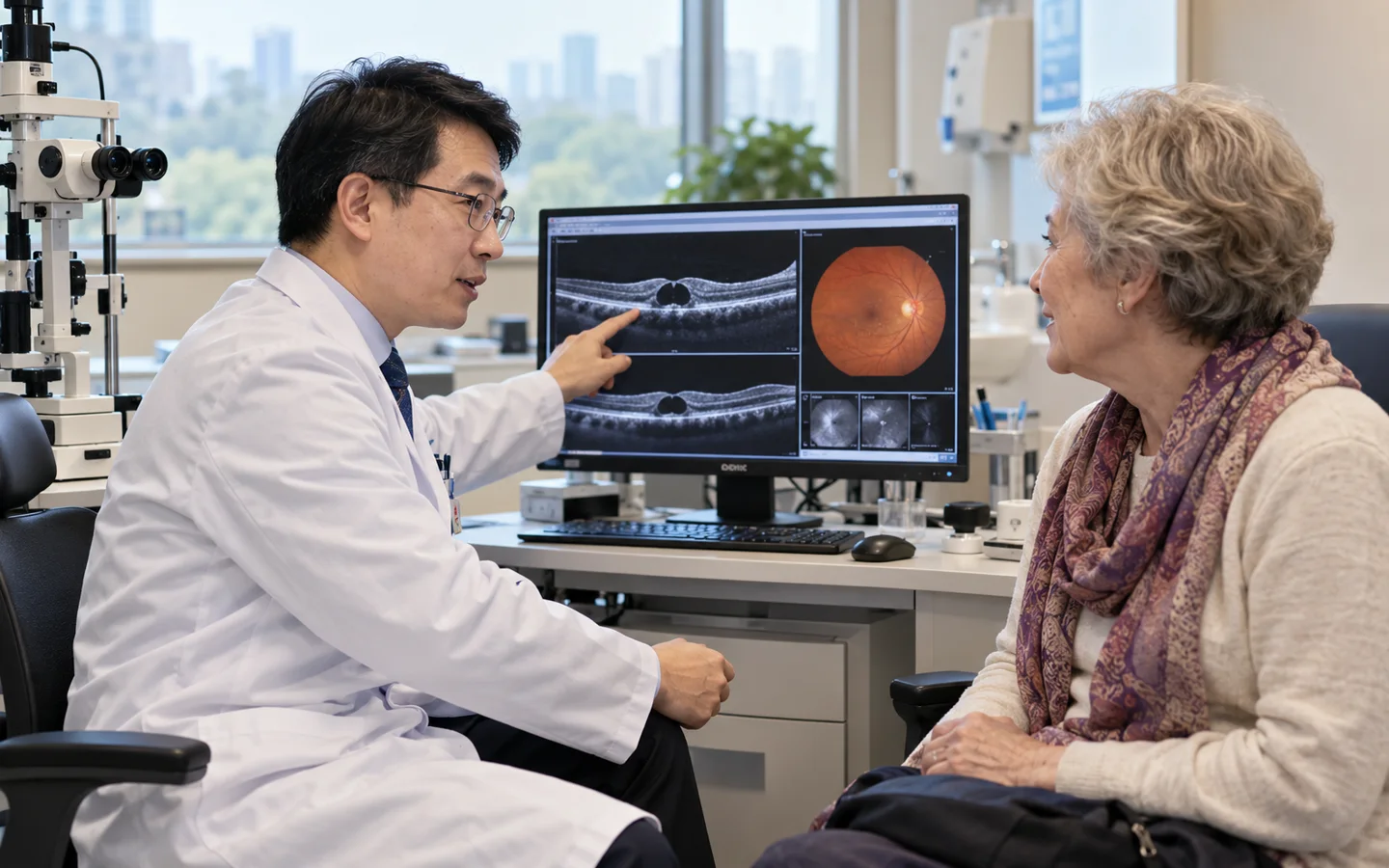
Chinese retina surgeon explaining macular hole and membrane surgery to an international older adult patient using OCT images

Medical records & cost enquiry

Macular Surgery: assessment and cost questions

For Macular Surgery, the budget depends on the proposed care and the hospital. A useful estimate needs to distinguish:

  • The macular condition and proposed operation
  • Additional eye procedures and materials
  • Positioning instructions and follow-up visits

Hospital medical fees, travel and our coordination services are separate. Any paid specialist review or coordination service is explained and agreed before you proceed.

Your next step

Start with your question

Tell us your diagnosis and what you need. Our free initial review checks the information and helps identify a suitable next step; it is not a specialist opinion or a hospital quotation.

Request a case-based estimate

Not ready to send records? Ask us first. Where hospital review is appropriate, we can help request an estimate. No travel commitment or mandatory proxy consultation.

Planning macular surgery in ChinaHospital review · individual costs · visit and follow-up

Plan the visit around retinal imaging, vision goals and surgical scope. Agree the assessment route before travel.

Records for the macular surgery review

Tell us what you already have: Serial macular OCT; Visual acuity and refraction; Amsler or distortion history. Start with a short summary; after first contact we explain which records the receiving team needs and how to share them.

Confirm the proposed scope and costs

Before asking for a personal estimate, clarify: The macular condition and proposed operation; Additional eye procedures and materials; Positioning instructions and follow-up visits. The receiving team confirms the proposed scope and hospital charges; coordination is agreed separately.

Visits and care after returning home

Discuss the specific macular problem and ask what postoperative positioning, eye reviews or other practical arrangements need confirmation. Tell us if you need interpretation or English-language documents, and confirm the relevant arrangements with the receiving team.

Macular surgery relieves traction or supports closure at the center of the retina

Vitrectomy removes the vitreous gel and fine membranes may be peeled from the macula. A gas bubble is often used for a macular hole.

The operation may improve distortion or protect central vision, but photoreceptor damage, duration and other retinal disease limit recovery.

Anatomical closure and visual recovery are different outcomes

Ask what OCT feature is being treated and what level of reading or distortion improvement is realistic.

Who may be considered?

Specialist review may help when the diagnosis, symptoms and previous treatment create a focused question about macular surgery.

  • A full-thickness macular hole affecting central vision.
  • A symptomatic epiretinal membrane causing distortion or reduced function.
  • Vitreomacular traction threatening or damaging the fovea.
  • A patient whose expected benefit outweighs cataract and retinal risks.

What the specialist team must confirm

Review serial macular OCT, visual acuity, distortion symptoms, lens status, peripheral retina, fellow-eye findings, duration and previous injections or retinal surgery.

Key points for this treatment
Targetshole membrane or traction
Imaginghigh-resolution macular OCT
Techniquevitrectomy with membrane work
Recoveryanatomy may improve before vision
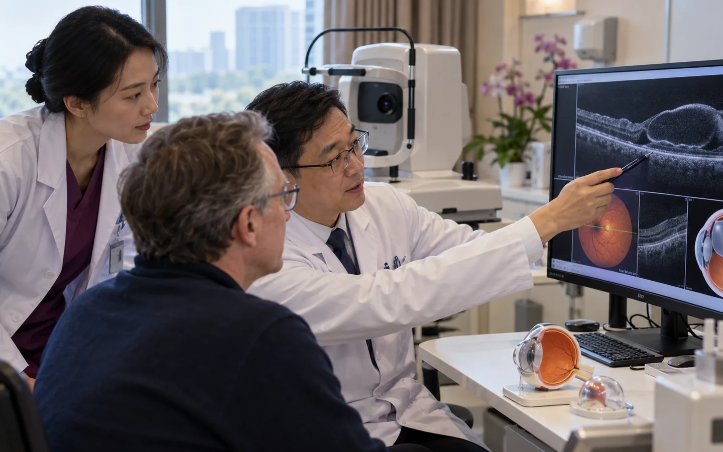
Chinese vitreoretinal team reviewing high resolution macular OCT gas bubble and membrane peeling plan
OCT defines the traction patternHole size, membrane anatomy, photoreceptor integrity and lens status influence technique and prognosis.

From OCT-defined traction to central-vision rehabilitation

Positioning and travel restrictions are discussed before surgery whenever an intraocular gas bubble may be used.

ConfirmLink symptoms to OCT anatomy
PlanChoose peel dye and tamponade
ReleaseRemove vitreous and traction
ObserveTrack closure structure and vision

Retinal anatomy can recover before visual function

Drops, positioning and restrictions depend on tamponade. Air travel and nitrous oxide are unsafe while a gas bubble remains.

OCT follows closure or contour; visual improvement can continue for months and may remain incomplete. Cataract often progresses after vitrectomy.
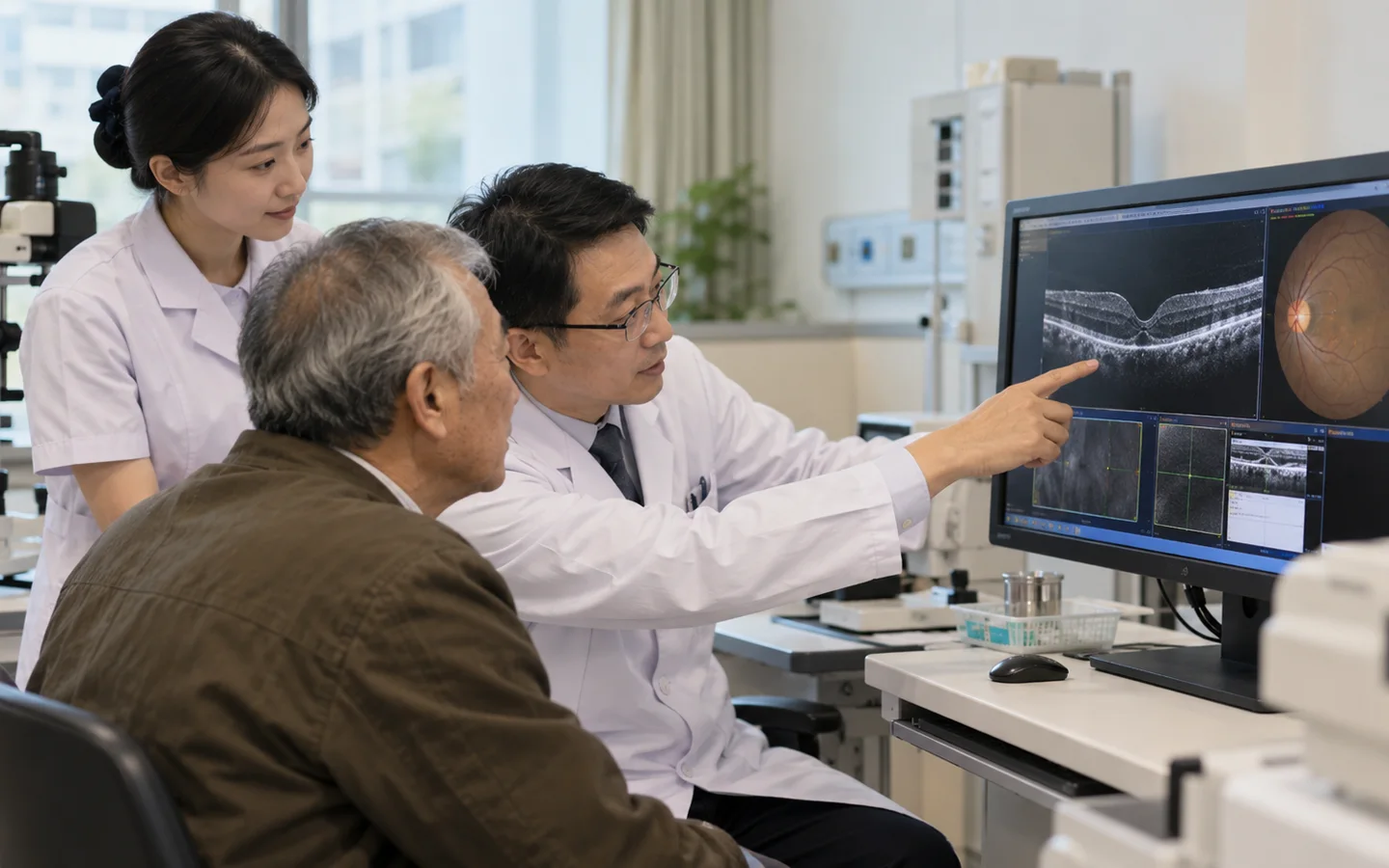
International older adult patient reviewing positioning visual distortion and OCT healing after macular surgery
Gas-bubble safety must be understoodThe patient should carry clear information about flying, altitude and anesthesia restrictions until the bubble is gone.
Hole closesContinue OCT and vision review
Distortion improvesAllow neural adaptation
Persistent tractionReassess anatomy and benefit
Cataract progressesPlan lens review when appropriate

Risks, limits and realistic expectations

Risks include cataract, retinal tear or detachment, infection, bleeding, pressure change, recurrent membrane or hole, visual-field defect and incomplete vision recovery.

Do not delay urgent local care

A curtain shadow, sudden shower of floaters, severe pain, rapid vision loss, marked redness or nausea after surgery requires urgent retinal assessment.

Before hospital review

Records for macular surgery assessment

A safe international review depends on dated source reports, original imaging and a complete treatment timeline—not a diagnosis label alone.

Serial macular OCT
Visual acuity and refraction
Amsler or distortion history
Dilated retinal examination
Lens and cataract status
Previous retinal injections or surgery
Fellow-eye findings
Medical and anticoagulation history

Tell us what you need

Ask about your care,
your hospital and your budget.

You can ask about suitability, an expert opinion, an appointment or the likely medical cost. If you are unsure, choose “Not sure — please advise”.

This enquiry is aboutMacular SurgeryNot sure — please advise

How a personal estimate is prepared

  1. Tell us about your case.Describe your diagnosis, main question and preferred city, if any.
  2. Share the relevant records.We explain what is needed and how to send it by WhatsApp or email.
  3. Request a hospital estimate.Where appropriate, we help request hospital review and a cost estimate. Any paid review is agreed first.

This is an enquiry, not an order or payment. Proxy consultation is not mandatory. Any service scope is agreed separately before you proceed.

Ask about Macular Surgery

A first enquiry is free. Email and permission to respond are required; the other details are optional. Any paid clinical review or coordination is discussed separately.

Included automatically so we know which procedure you are asking about.
A preference, not a confirmed appointment.
Please do not send passport numbers, card details or full medical files in this first enquiry. We will explain which records are needed next.

Send a short summary first. This is an enquiry, not an order, payment or confirmed appointment.

No booking or payment is made by sending this enquiry.
Editorial transparency

Medical sources

Patient information is based on established government and professional guidance. Content updated 5 October 2026. This is patient information, not an individual clinical assessment.