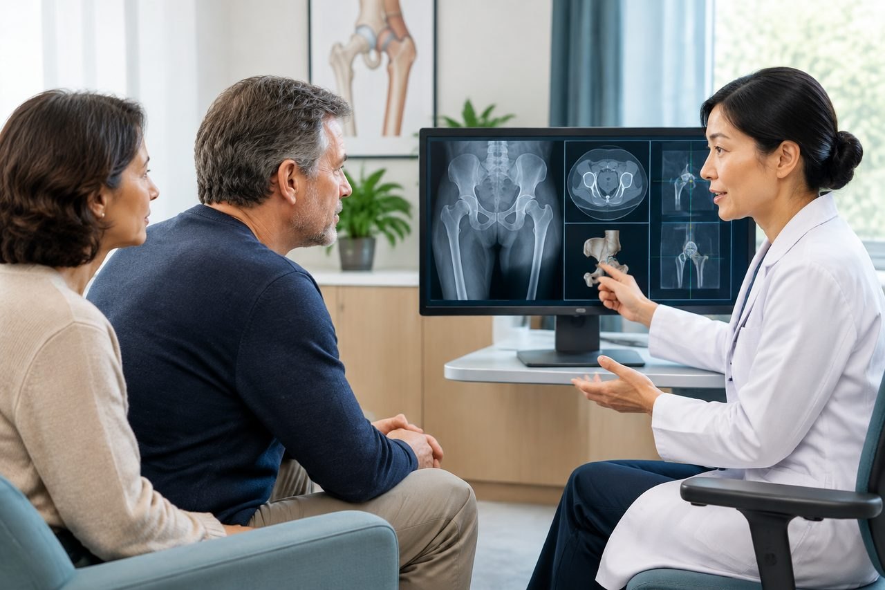
Why the imaging request differs from one team to the next
Robotic hip replacement is not one identical operation. It is a group of techniques in which navigation or robotic tools help the surgeon position components and follow a preoperative plan. The American Academy of Orthopaedic Surgeons describes robotic-assisted joint replacement as a tool that assists planning and surgery while the surgeon remains responsible for the operation. Because the toolset varies, the images needed to feed that plan also vary.
Three things change the request. First, the condition being treated: osteoarthritis, avascular necrosis, a previous fracture, dysplasia or a failed earlier implant each need different information. Second, the surgical approach: a surgeon planning an anterior approach may want different views from one planning a posterior approach. Third, the platform: some systems build a three-dimensional model from a CT scan, while others rely mainly on intraoperative registration with plain films. None of these is automatically better; they are different planning routes.
This is why a generic list of scans is not enough. The useful question is not 'what imaging does robotic hip surgery need' but 'what does this specific team need for this specific hip'.
What each type of imaging actually contributes
A plain radiograph, usually an anteroposterior view of the pelvis plus a lateral view of the affected hip, remains the baseline. It shows the joint space, the shape of the socket, the position of the femoral head and any deformity. Many surgeons also ask for a weight-bearing or standing view, because a hip looks different when you are lying down. If you have older films, they are still useful for showing how the joint has changed over time.
A CT scan of the pelvis and hip gives three-dimensional information that a plain film cannot. It helps measure version, assess bone stock and detect cysts or defects that affect component choice. Some robotic workflows use CT data to create a patient-specific plan before the operation. A low-dose protocol may be requested to limit radiation, but whether that is available and appropriate is a decision for the treating radiologist and surgeon, not something to arrange on your own.
An MRI is not routine for hip replacement planning. It is more often used when the diagnosis is unclear, when a soft-tissue problem such as a labral tear is suspected, or when avascular necrosis needs staging. A bone scan or SPECT may be requested in selected cases. Each of these answers a different question, so sending everything 'just in case' can slow review rather than speed it up.
Blood tests, an ECG and a chest radiograph are usually part of fitness-for-surgery assessment rather than implant planning. They belong to the anaesthetic and medical workup, which the hospital will define separately.
A labelled planning example: how the request changes
This is an illustration of reasoning, not medical advice. Suppose a 62-year-old with primary osteoarthritis and no previous hip surgery asks about robotic hip replacement. A team using a CT-based plan may ask for a recent standing AP pelvis, a lateral hip view and a thin-slice CT of the affected hip. A team using an imageless system may ask only for good-quality plain films and may take additional registration images in the operating theatre.
Now change one detail: the same patient had a previous hip fracture fixed with screws years ago. The hardware can distort CT images and obscure anatomy, so the team may ask for the original operative notes, the implant details and additional views. Change another detail: the patient has severe dysplasia. The surgeon may want a CT to measure the socket and plan cup position, and may also want a standing full-length view to assess leg length.
The lesson is that the imaging list follows the clinical question. When you send records, include the report as well as the images, and note the date of each study. A report from a different hospital is still useful, but the receiving team may want to review the actual image files rather than rely on the text alone.
How to send imaging to a team in China
Most hospitals prefer DICOM files on a disc or a secure download link rather than photographs of a screen. Photographs lose detail and cannot be measured. If you only have printed films, ask the original imaging centre whether they can export the digital files. If you have a radiology report in another language, a short summary in English or Chinese helps the first review, but the original report should travel with it.
Before sending anything, ask the receiving team three questions: which views and which modality they want, how recent the study should be, and whether they need the raw DICOM files or will accept a report first. The answers change what you request from your local imaging centre. Do not order a new CT or MRI on your own initiative; a scan done in the wrong protocol may need to be repeated.
It also helps to say what you already have. If you have a CT from six months ago and a plain film from last month, list both with dates. The team can then tell you whether the older study is still usable or whether a new one is needed. This is faster than sending a large archive and waiting for someone to sort through it.
- A one-page summary: age, main hip problem, previous hip surgery, current walking ability and main question.
- Recent plain radiographs with dates and reports.
- Any CT, MRI or bone scan already done, with reports.
- Previous operative notes and implant details if you have had hip surgery before.
- A list of current medicines and relevant allergies.
What imaging cannot tell you before you travel
Imaging supports planning, but it does not confirm that robotic hip replacement is suitable for you, that a particular hospital will accept your case, or that a specific robotic platform is available. Those are clinical and administrative decisions made by the treating team after reviewing your records and, often, examining you in person. A records-based opinion can clarify whether the approach is worth pursuing, but it is not final procedural clearance.
It is also worth separating three things that are often confused: the implant, the robotic platform and the surgeon. A hospital may have a robotic system but use it for selected cases only. A surgeon may be experienced with hip replacement but use robotic assistance for a subset of patients. The implant brand is a separate choice again. Ask which of these applies to your case rather than assuming that the presence of a robot determines the plan.
If your symptoms are worsening, or you have new pain, fever, inability to bear weight or a recent fall, seek local medical assessment first. Planning an overseas operation should not delay urgent care.
Questions that change the next step
The answers to a small number of questions determine whether you can proceed with a records-based review or whether you need more imaging first. Ask the receiving team directly, and keep the answers in writing.
Which imaging do you need for planning, and in what format? Is a CT required, or will plain films plus intraoperative registration be enough? How recent must the studies be? Do you need weight-bearing views? If I have had previous hip surgery, do you need the old operative notes and implant details? Will the imaging be repeated in China, and if so, what does that involve? These are practical questions, not a test of the team.
You can also ask whether a records-based opinion is possible before you travel, and what it can and cannot establish. A remote review can help decide whether robotic hip replacement is a reasonable route to explore, but it does not replace an in-person assessment or guarantee hospital acceptance.
A practical next step
Start with a short summary rather than a complete archive. ChinaSpecialistCare's free initial case review checks the available diagnosis, records and your main question, identifies missing information and suggests the relevant next step. It is not a diagnosis or a promise of acceptance. If a records-based specialist opinion is useful, that can be discussed separately; it is optional and not a prerequisite for every appointment.
Send the summary through the enquiry form, email or WhatsApp. After first contact, the team can explain how to share imaging securely. Do not send passport numbers, card details or a full medical archive at the first step.
Sources & scope of this guide
References and official service information relevant to this guide.
This is general planning information and has not been individually reviewed by a doctor. Medical decisions and personal treatment advice come from your treating clinicians.