Home / ENT (Ear, Nose & Throat) / Endoscopic Skull Base Surgery
ENT patient guide · 鼻内镜颅底手术

Endoscopic Skull Base Surgery in China

Considering endoscopic skull base surgery in China? Start with tumour anatomy, prior treatment and specialist-team assessment. This guide helps you identify the relevant records, questions for the receiving team and the scope of an individual estimate before a visit is agreed.

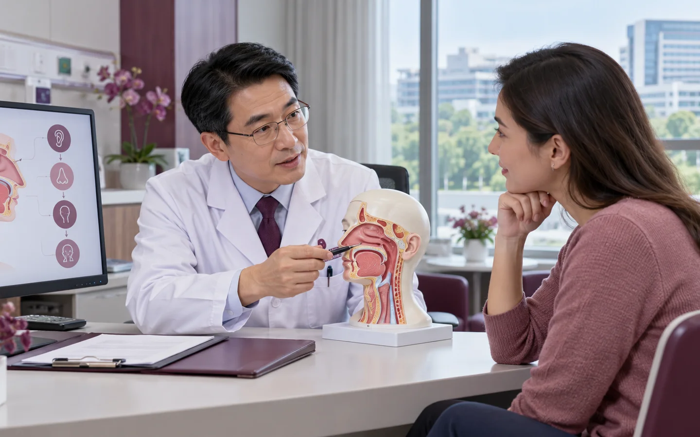
Chinese ENT specialist explaining endoscopic skull-base approach and team planning using a head model to a fully clothed international patient

Medical cost reference

Indicative · not a quotation

Endoscopic Skull Base Surgery costs in China

US$14,500–42,000

Original USD reference

Other currencies are approximate equivalents, not a quote or payment rate. Reference FX: ECB · 2 October 2026. Local snapshot, not live.

Endoscopic skull-base surgery; lesion location, reconstruction and admission affect costs.

This published reference range is a starting point for planning, not a fixed price or a China-wide average. Cities, public and private hospitals, procedure details and individual care needs affect costs. The hospital must confirm what is included and provide an estimate after reviewing your case.

  • Lesion location and extent
  • Reconstruction and specialist-team requirements
  • Admission and subsequent follow-up

Hospital medical fees, travel and our coordination services are separate. Any paid specialist review or coordination service is explained and agreed before you proceed.

Your next step

Start with your question

Tell us your diagnosis and what you need. Our free initial review checks the information and helps identify a suitable next step; it is not a specialist opinion or a hospital quotation.

Request a case-based estimate

Not ready to send records? Ask us first. Where hospital review is appropriate, we can help request an estimate. No travel commitment or mandatory proxy consultation.

Planning endoscopic skull base surgery in ChinaHospital review · individual costs · visit and follow-up

Plan the visit around tumour anatomy, prior treatment and specialist-team assessment. Agree the assessment route before travel.

Records for the endoscopic skull base surgery review

Tell us what you already have: All MRI DICOM studies; Fine-cut skull-base CT DICOM; Vascular imaging if performed. Start with a short summary; after first contact we explain which records the receiving team needs and how to share them.

Confirm the proposed scope and costs

Review the published reference below with the receiving team. Confirm the particular procedure, hospital and items included in your own estimate; the reference is not a confirmed quotation.

Visits and care after returning home

Ask which surgical and other specialists need to assess the case and how their proposed care and follow-up fit together. Tell us if you need interpretation or English-language documents, and confirm the relevant arrangements with the receiving team.

Endoscopic Skull Base Surgery: diagnosis, function and long-term planning

Endoscopic Skull Base Surgery is considered when a selected pituitary, sinonasal or skull-base lesion can be reached endonasally with acceptable neurovascular and reconstructive risk.

The safest approach depends on tumor compartment, pathology, carotid and optic relationships, cavernous-sinus involvement, hormonal status and the ability to create a watertight closure.

Who may be considered?

Specialist review may help when a central skull-base lesion requires comparison of endoscopic, open, radiation or observation strategies.

  • Selected pituitary and midline skull-base tumors.
  • Sinonasal tumors extending to the skull base.
  • Cerebrospinal-fluid leaks requiring repair.
  • A patient evaluated by coordinated skull-base specialists.

What the specialist team must confirm

Review contrast MRI and fine-cut CT DICOM, vascular imaging when relevant, formal visual fields, pituitary hormones, nasal endoscopy, pathology, neurologic examination and previous surgery or radiation.

Key points for this treatment

Approachoften through nose and sinuses
TeamENT plus neurosurgery
PlanningMRI CT vessels and pathology
Prioritysafe removal and reliable closure
Chinese multidisciplinary ENT team reviewing fused MRI CT vascular optic nerve and closure-reconstruction planning
Access and closure are planned togetherResection goals must be balanced with carotid, optic, cranial-nerve, hormonal and cerebrospinal-fluid leak risks.

From multidisciplinary mapping to staged skull-base surveillance

The plan should define resection limits, pathology contingencies and postoperative endocrine, visual and nasal care.

MapFuse tumor bone vessels and nerves
CoordinateAlign ENT neurosurgery and endocrine plans
AccessUse the safest corridor
CloseReconstruct and monitor leak risk

Neurologic, endocrine and nasal recovery are monitored together

Early care checks vision, neurologic status, sodium and hormones, nasal bleeding, infection and cerebrospinal-fluid leakage.

MRI, pathology-specific care, endocrine replacement, nasal endoscopy and rehabilitation continue according to the lesion and resection result.

Fully clothed international patient completing vision hormone nasal healing and neurologic follow up after skull-base surgery with a Chinese ENT clinician
Several systems recover on different timelinesA shared follow-up plan prevents visual, hormonal or closure problems from falling between specialties.
Planned resectionContinue pathology-led surveillance
Residual tumorConsider observation radiation or staged care
Hormone deficitStart endocrine replacement
CSF leak concernAssess and repair urgently

Risks, limits and realistic expectations

Risks include cerebrospinal-fluid leak, meningitis, bleeding, carotid injury, visual loss, cranial-nerve injury, stroke, hormonal disturbance, smell loss and need for further treatment.

Do not delay urgent local care

Clear salty nasal drainage, severe headache, fever, neck stiffness, visual decline, confusion, marked thirst or reduced consciousness needs emergency assessment.

Before hospital review

Records for endoscopic skull base surgery assessment

A safe international review depends on dated source reports, original imaging and a complete treatment timeline—not a diagnosis label alone.

All MRI DICOM studies
Fine-cut skull-base CT DICOM
Vascular imaging if performed
Visual fields and acuity
Pituitary hormone results
Nasal endoscopy findings
Pathology and molecular results
Previous surgery and radiotherapy records

Tell us what you need

Ask about your care,
your hospital and your budget.

You can ask about suitability, an expert opinion, an appointment or the likely medical cost. If you are unsure, choose “Not sure — please advise”.

This enquiry is aboutEndoscopic Skull Base SurgeryNot sure — please advise

How a personal estimate is prepared

  1. Tell us about your case.Describe your diagnosis, main question and preferred city, if any.
  2. Share the relevant records.We explain what is needed and how to send it by WhatsApp or email.
  3. Request a hospital estimate.Where appropriate, we help request hospital review and a cost estimate. Any paid review is agreed first.

This is an enquiry, not an order or payment. Proxy consultation is not mandatory. Any service scope is agreed separately before you proceed.

Ask about Endoscopic Skull Base Surgery

A first enquiry is free. Email and permission to respond are required; the other details are optional. Any paid clinical review or coordination is discussed separately.

Included automatically so we know which procedure you are asking about.
A preference, not a confirmed appointment.
Please do not send passport numbers, card details or full medical files in this first enquiry. We will explain which records are needed next.

Send a short summary first. This is an enquiry, not an order, payment or confirmed appointment.

No booking or payment is made by sending this enquiry.
Editorial transparency

Medical sources

Patient information is based on established government and professional guidance. Content updated 5 October 2026. This is patient information, not an individual clinical assessment.