
Corneal transplantation can replace only the diseased layer or the full cornea
Penetrating keratoplasty replaces full thickness, while endothelial or anterior lamellar procedures target selected layers.
The diagnosis, scarring depth, endothelial health, lens, retina, glaucoma and previous infection determine which transplant is appropriate and how much vision can improve.
Ask which layer is failing, why that transplant type is chosen and whether donor tissue access affects timing.
Who may be considered?
Specialist review may help when the diagnosis, symptoms and previous treatment create a focused question about corneal transplant.
- Central scarring or irregularity not adequately treated by lenses or laser.
- Endothelial failure causing persistent corneal swelling.
- Advanced keratoconus or dystrophy unsuitable for lesser treatment.
- A patient able to use long-term drops and attend rejection surveillance.
What the specialist team must confirm
Review slit-lamp findings, corneal topography and tomography, pachymetry, endothelial-cell status, infection history, glaucoma, lens and retinal potential and previous grafts.
Key points for this treatment
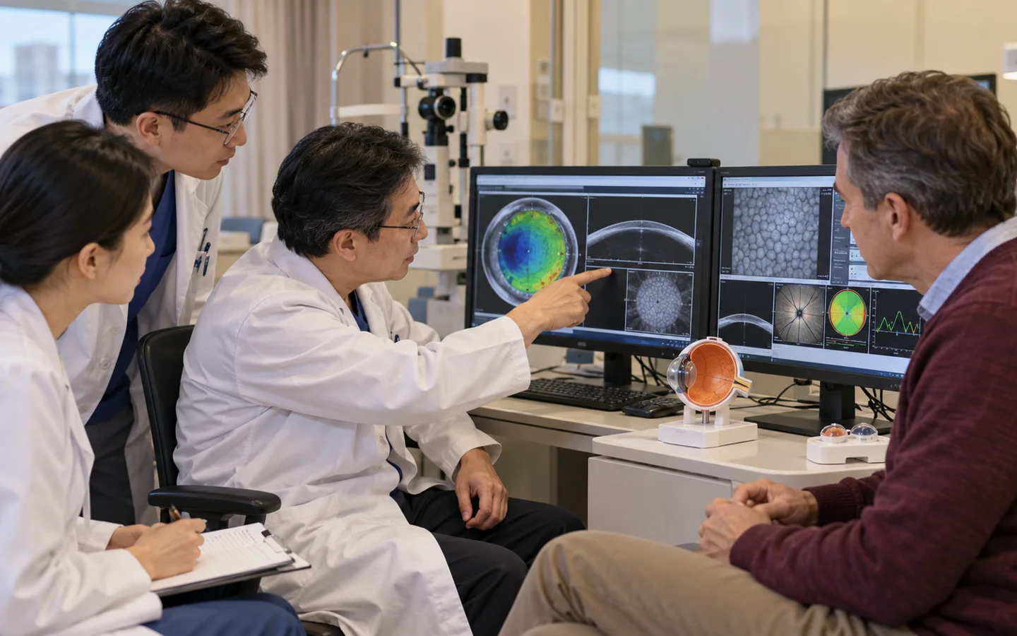
From layer diagnosis to long-term graft protection
Visual rehabilitation may include suture adjustment, glasses, contact lenses or later cataract treatment after graft stability.
Graft clarity depends on medication and rapid response to symptoms
Drops reduce inflammation and rejection. Vision may fluctuate while swelling resolves or sutures and corneal shape change.
Long-term checks monitor rejection, infection, pressure, astigmatism, endothelial health and the rest of the eye.
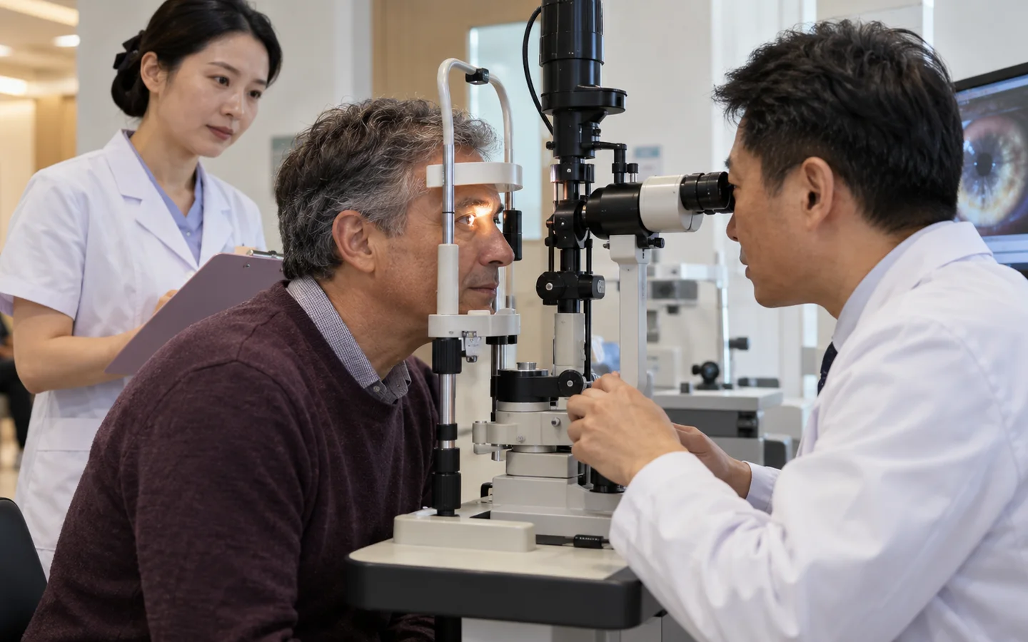
Risks, limits and realistic expectations
Risks include rejection, infection, bleeding, glaucoma, cataract, wound opening, retinal problems, irregular astigmatism, graft failure and need for repeat transplantation.
New pain, redness, light sensitivity, cloudy vision, discharge or sudden vision loss after transplant requires urgent ophthalmic assessment.