
Home / Gastroenterology / Endoscopic Submucosal Dissection (ESD)
Gastroenterology patient guide · 内镜黏膜下剥离术

Endoscopic Submucosal Dissection in China

Considering endoscopic submucosal dissection in China? Start with lesion reports, pathology and endoscopic treatment scope. This guide helps you identify the relevant records, questions for the receiving team and the scope of an individual estimate before a visit is agreed.

Chinese advanced endoscopist explaining ESD and pathology margins to an international patient

Medical records & cost enquiry

Endoscopic Submucosal Dissection: assessment and cost questions

For Endoscopic Submucosal Dissection (ESD), the budget depends on the proposed care and the hospital. A useful estimate needs to distinguish:

  • Lesion location, size and number
  • Endoscopic resection and pathology scope
  • Admission and subsequent surveillance

Hospital medical fees, travel and our coordination services are separate. Any paid specialist review or coordination service is explained and agreed before you proceed.

Your next step

Start with your question

Tell us your diagnosis and what you need. Our free initial review checks the information and helps identify a suitable next step; it is not a specialist opinion or a hospital quotation.

Request a case-based estimate

Not ready to send records? Ask us first. Where hospital review is appropriate, we can help request an estimate. No travel commitment or mandatory proxy consultation.

Planning endoscopic submucosal dissection in ChinaHospital review · individual costs · visit and follow-up

Plan the visit around lesion reports, pathology and endoscopic treatment scope. Agree the assessment route before travel.

Records for the endoscopic submucosal dissection review

Tell us what you already have: High-quality endoscopy images and report; Biopsy pathology slides or blocks; EUS CT or MRI staging when performed. Start with a short summary; after first contact we explain which records the receiving team needs and how to share them.

Confirm the proposed scope and costs

Before asking for a personal estimate, clarify: Lesion location, size and number; Endoscopic resection and pathology scope; Admission and subsequent surveillance. The receiving team confirms the proposed scope and hospital charges; coordination is agreed separately.

Visits and care after returning home

Ask which source images and pathology the endoscopist needs, and whether the proposed estimate includes specimen review and later surveillance. Tell us if you need interpretation or English-language documents, and confirm the relevant arrangements with the receiving team.

ESD separates a superficial lesion from the deeper wall in one piece

The endoscopist marks the lesion, injects beneath it, makes a mucosal incision and carefully dissects the submucosal layer. En-bloc removal helps pathologists assess margins and depth.

Suitability depends on size, location, fibrosis, ulceration and the estimated chance of deep invasion or lymph-node spread. ESD is technically demanding and not equivalent to routine polypectomy.

The final pathology decides whether ESD was curative

A completely removed lesion may still need surgery if invasion, lymphatic or vascular involvement or other high-risk features exceed accepted criteria.

Who may be considered?

Specialist review may help when the diagnosis, symptoms and previous treatment create a focused question about endoscopic submucosal dissection.

  • Selected early esophageal or gastric cancer confined to superficial layers.
  • A large colorectal lesion suitable for organ-preserving en-bloc resection.
  • A lesion where piecemeal removal would impair staging.
  • A patient assessed in a centre with advanced endoscopic and surgical backup.

What the specialist team must confirm

Review includes high-definition endoscopy, lesion morphology, targeted biopsy, chromoendoscopy or magnification, EUS or cross-sectional staging when appropriate, prior attempted resection, anticoagulation, comorbidity and access to pathology and emergency surgery.

Key points for this treatment
Goalremove selected superficial lesions en bloc
Sitesesophagus stomach colon and rectum
Evidenceendoscopy imaging biopsy and staging
Key resultmargin depth and pathology
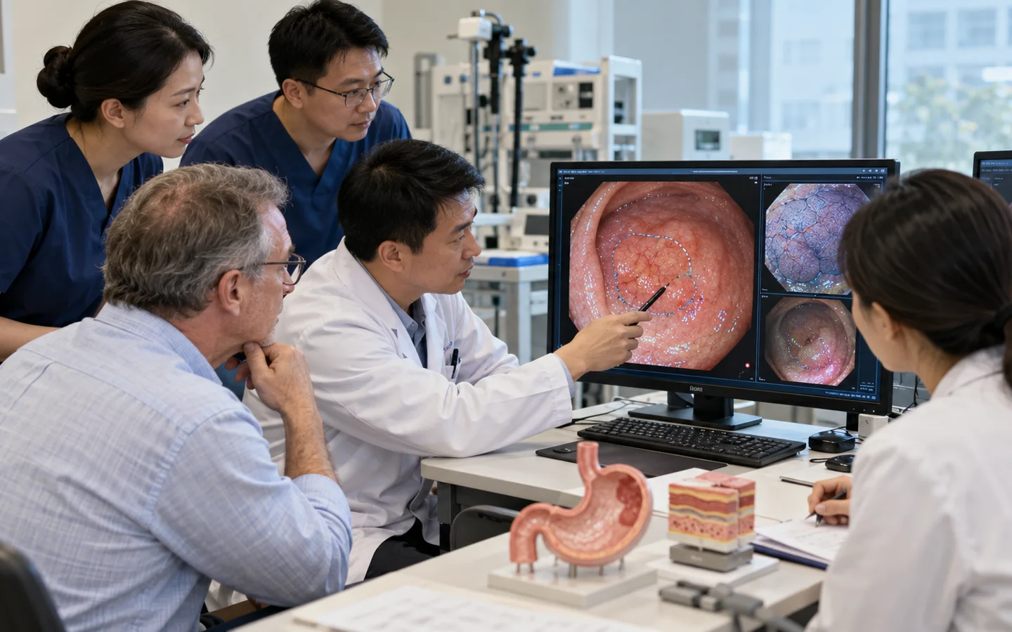
Chinese ESD team reviewing magnified endoscopy lesion boundaries and submucosal dissection plan
Surface pattern and depth estimate guide selectionThe team maps lesion borders and predicts invasion before deciding whether endoscopic removal can be both complete and oncologically adequate.

From optical diagnosis to pathology-defined cure

ESD is selected when complete endoscopic staging and organ preservation are plausible without delaying necessary surgery.

CharacterizeMap margins and invasion risk
LiftInject and open the mucosal plane
DissectRemove lesion en bloc
StageReview depth margins and risk features

Healing and pathology both require follow-up

Aftercare varies by organ and may include fasting, acid suppression, observation and staged diet. Bleeding or perforation can occur during or after the procedure.

The pathology report should state size, histology, depth, margins and lymphovascular invasion. Surveillance or additional surgery follows that risk assessment.

International patient reviewing diet healing and final pathology after endoscopic submucosal dissection
En-bloc tissue provides the answerOrientation and expert pathology allow the multidisciplinary team to judge whether the resection meets curative criteria.
Curative pathologyEnter endoscopic surveillance
High-risk featureDiscuss additional surgery
Delayed bleedingTreat promptly
Narrowing riskPlan symptom and endoscopic review

Risks, limits and realistic expectations

Risks include bleeding, perforation, post-procedure pain, infection, stricture and need for emergency surgery. ESD does not treat lymph-node disease and may be noncurative on final pathology.

Do not delay urgent local care

Severe chest or abdominal pain, fever, vomiting blood, black stool, fainting or breathing difficulty after ESD requires urgent local care.

Before hospital review

Records for endoscopic submucosal dissection assessment

A safe international review depends on dated source reports, original imaging and a complete treatment timeline—not a diagnosis label alone.

High-quality endoscopy images and report
Biopsy pathology slides or blocks
EUS CT or MRI staging when performed
Prior resection or biopsy procedure notes
Anticoagulation and medicine list
Cardiopulmonary and anesthesia assessment
Surgical consultation when relevant
Final pathology criteria and surveillance plan

Tell us what you need

Ask about your care,
your hospital and your budget.

You can ask about suitability, an expert opinion, an appointment or the likely medical cost. If you are unsure, choose “Not sure — please advise”.

This enquiry is aboutEndoscopic Submucosal Dissection (ESD)Not sure — please advise

How a personal estimate is prepared

  1. Tell us about your case.Describe your diagnosis, main question and preferred city, if any.
  2. Share the relevant records.We explain what is needed and how to send it by WhatsApp or email.
  3. Request a hospital estimate.Where appropriate, we help request hospital review and a cost estimate. Any paid review is agreed first.

This is an enquiry, not an order or payment. Proxy consultation is not mandatory. Any service scope is agreed separately before you proceed.

Ask about Endoscopic Submucosal Dissection (ESD)

A first enquiry is free. Email and permission to respond are required; the other details are optional. Any paid clinical review or coordination is discussed separately.

Included automatically so we know which procedure you are asking about.
A preference, not a confirmed appointment.
Please do not send passport numbers, card details or full medical files in this first enquiry. We will explain which records are needed next.

Send a short summary first. This is an enquiry, not an order, payment or confirmed appointment.

No booking or payment is made by sending this enquiry.
Editorial transparency

Medical sources

Patient information is based on established government and professional guidance. Content updated 5 October 2026. This is patient information, not an individual clinical assessment.