Lengthening is a biological process, not one operation
Limb lengthening uses distraction osteogenesis: bone is divided in a controlled osteotomy, then separated gradually so new bone forms in the gap. It may treat a clinically important congenital or acquired limb-length difference, angular deformity or bone loss after trauma or infection.
The procedure also stretches muscles, nerves, vessels and skin, which limits speed and total correction. Shoe lifts, guided growth, acute correction, shortening of the longer limb or accepting a small difference may be safer alternatives. This page addresses medically indicated reconstruction, not cosmetic height increase.
Adjustment, pin or wound care, joint exercises, weight-bearing rules and serial X-rays must continue after the patient leaves hospital. A local team must be arranged before international travel.
Who may be considered?
A limb-reconstruction team may consider gradual correction for:.
- A meaningful discrepancy causing gait, pelvic or functional problems.
- Congenital shortening such as selected femoral or tibial deficiencies.
- Post-traumatic shortening, malunion or bone loss.
- Deformity after infection, growth-plate injury or previous surgery.
- A patient and support system able to manage a long, demanding treatment course.
What the hospital needs to assess
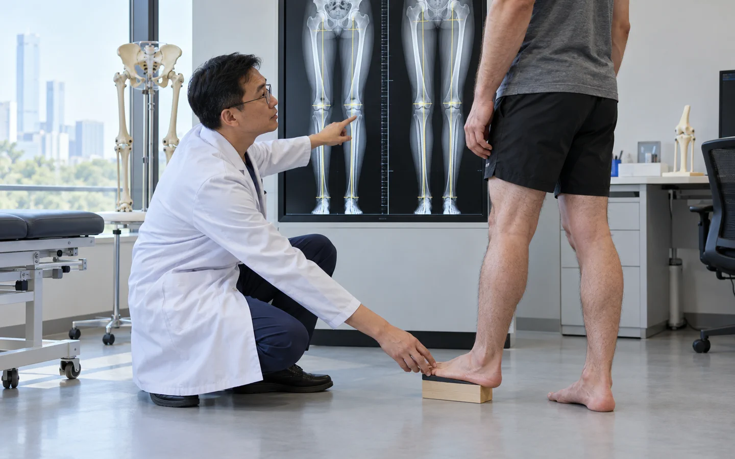
Standing full-length radiographs with calibration measure length and mechanical axis. Clinical blocks separate true limb-length difference from pelvic or joint contracture. CT may assess rotation or bone anatomy. The team examines hip, knee and ankle motion, nerve status, scars, infection history, bone health, maturity and psychosocial readiness.
Key points for this treatment
Internal nails and external fixation
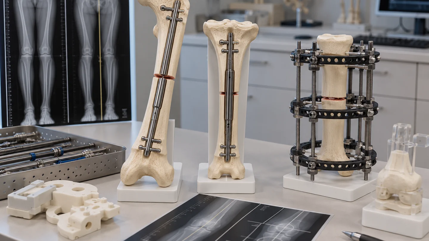
After osteotomy, an internal motorised nail can gradually lengthen selected bones when canal size, growth status and weight limits are suitable. External fixators connect bone to an outside frame with pins or wires and can correct complex multiplanar deformity, translation and rotation.
Following a latency period, the device is adjusted in small daily increments. X-rays guide changes in rate if regenerate bone is too weak or consolidating too early. When the target is reached, the new bone must harden before full loading or frame removal.
Hospital stay and recovery
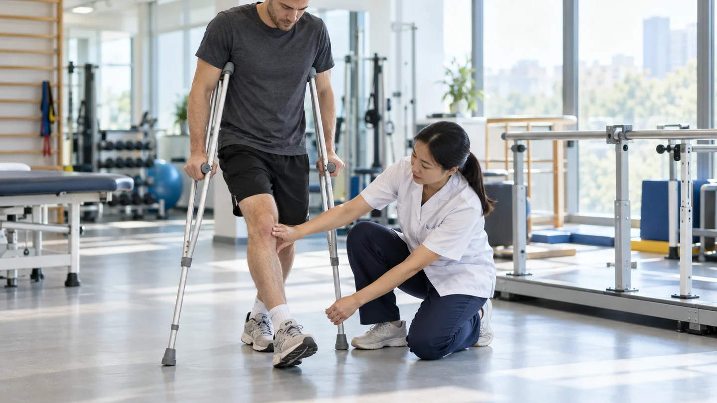
Physiotherapy begins early and is often daily. Maintaining knee and ankle motion is essential because soft tissues can tighten as length increases. Crutches or a walker manage device-specific weight-bearing; nutrition and vitamin status may be reviewed.
Treatment can last many months. Serial radiographs monitor regenerate bone and alignment. Hardware may later be removed. International patients need rapid access to the operating team for device malfunction, nerve symptoms, joint contracture, pin infection or unexpected pain.
Risks and realistic expectations
Risks include pin-site or deep infection, joint stiffness or contracture, nerve or vessel injury, pain, poor regenerate bone, premature consolidation, delayed union or non-union, fracture, malalignment, device failure, blood clot and additional surgery. The target may need to be reduced to protect function.
New numbness or weakness, severe pain during adjustment, a cold or pale foot, fever, spreading pin-site redness, device breakage, sudden deformity, calf swelling, chest pain or breathlessness requires urgent review.