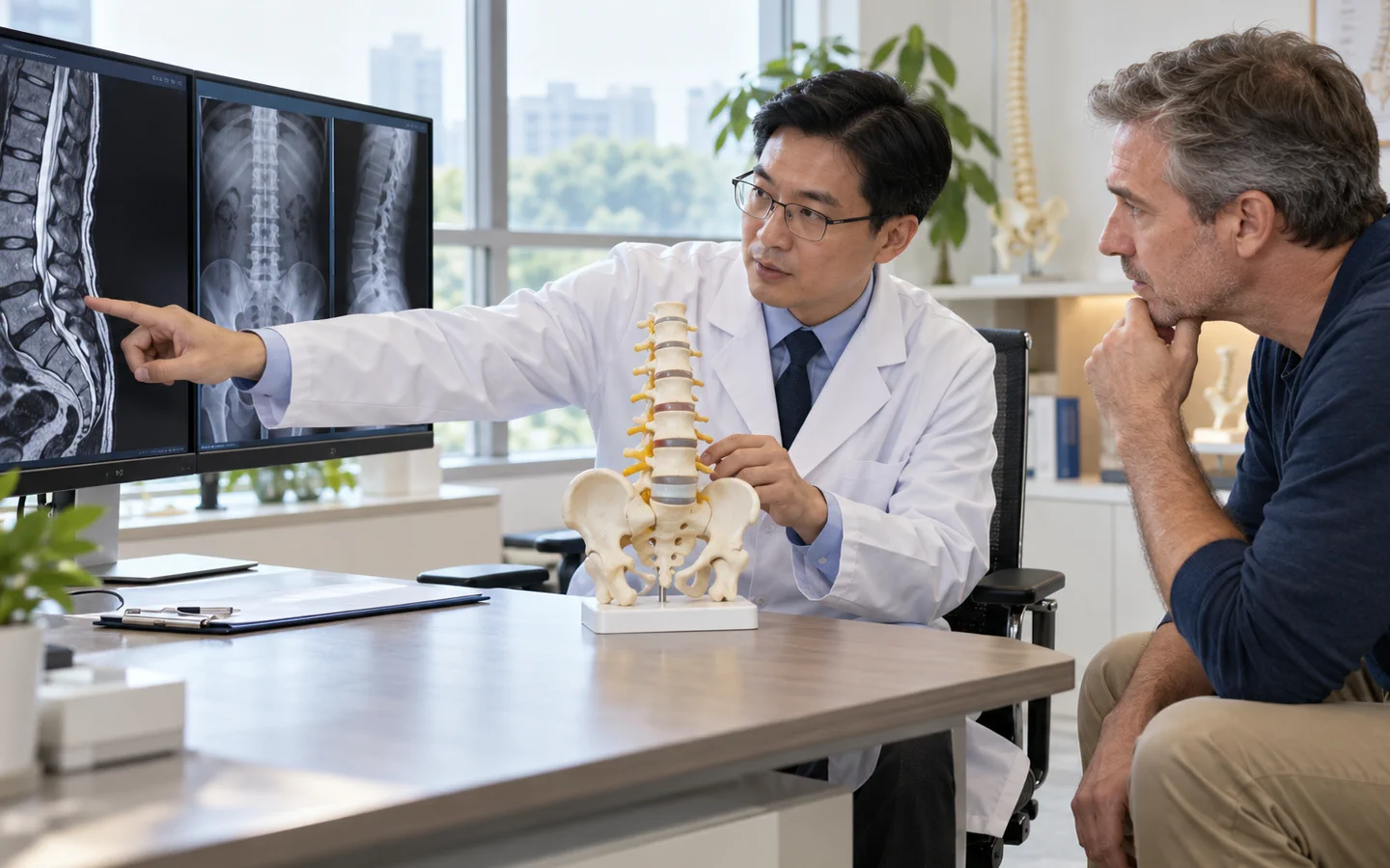
What lumbar fusion does—and does not do
Lumbar fusion aims to stop motion between selected lower-back vertebrae by creating a solid bridge of bone. It may be used to stabilise spondylolisthesis, support deformity correction, reconstruct a damaged segment or maintain stability after a necessary decompression.
Low-back pain has many possible sources. A worn disc on MRI does not by itself prove that fusion will help. The surgeon should explain the diagnosis, why the painful or unstable level is believed to be responsible, and why non-surgical care or decompression alone is insufficient.
Evidence and recommendations differ by diagnosis. The decision should be based on instability, deformity, neurological compression or another defined structural problem—not a scan label alone.
Who may be considered?
Lumbar fusion may be considered in carefully selected patients with:.
- Symptomatic spondylolisthesis or instability that matches imaging.
- Stenosis requiring decompression when instability is present or would be created.
- Adult deformity with imbalance and disabling symptoms.
- Fracture, tumour, infection or severe structural damage needing stabilisation.
- Non-union, implant failure or adjacent-level disease after previous surgery.
What the hospital needs to assess
Standing X-rays show alignment and slip under load; flexion-extension views may assess movement. MRI shows stenosis and nerves, while CT defines bone, facets and previous fusion. The team reviews symptoms in the back versus legs, walking tolerance, neurological findings, bone density, nicotine exposure and metabolic health.
Key points for this treatment
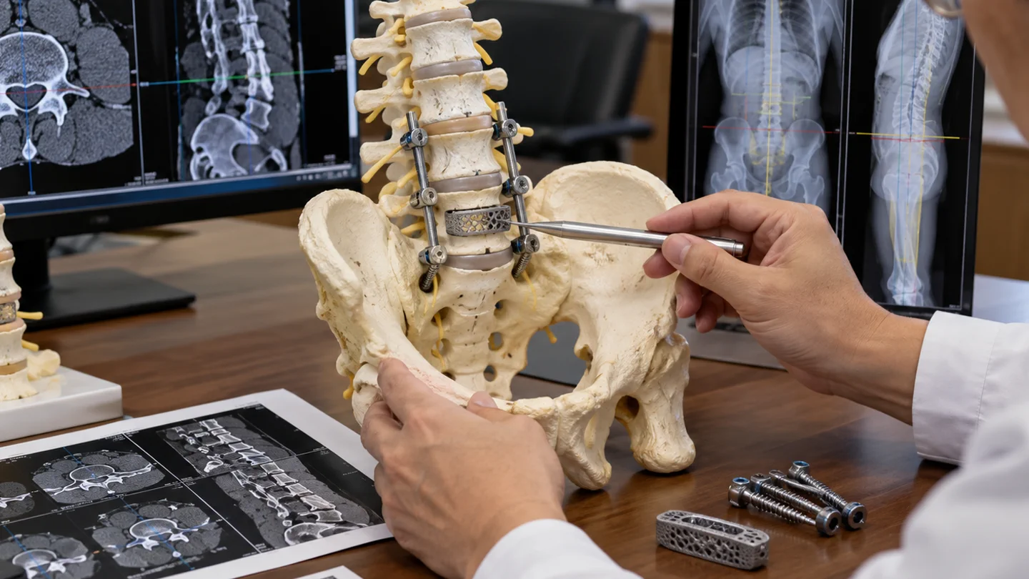
Common lumbar fusion approaches
Posterior operations include TLIF or PLIF, which access the disc space from the back while permitting nerve decompression. ALIF approaches from the abdomen; lateral approaches reach selected levels from the side. A posterolateral fusion places graft along the back of the spine and may be combined with interbody fusion.
Cages can restore disc height and hold graft; pedicle screws and rods stabilise the level. More than one route may be combined for complex alignment, but a larger construct also increases exposure and recovery demands.
Hospital stay and recovery
Early walking helps reduce immobility complications, but lifting, twisting and prolonged positions are progressed cautiously. Leg symptoms may improve at a different pace from mechanical back pain, and nerves compressed for a long time may not recover fully.
Fusion develops over months. Follow-up includes symptoms, neurological examination and imaging. Nicotine avoidance, nutrition and control of diabetes are important modifiable factors. International travel requires surgeon clearance and a local follow-up plan.

Risks and realistic expectations
Risks include infection, bleeding, blood clots, medical complications, nerve injury, spinal-fluid leak, persistent symptoms, non-union, cage migration, screw or rod failure and adjacent-level degeneration. Anterior and lateral approaches add risks to blood vessels, abdominal organs, nerves or muscles specific to the route.
New leg weakness, saddle numbness, loss of bladder or bowel control, fever, wound drainage, chest pain, breathlessness or severe escalating pain requires urgent assessment.