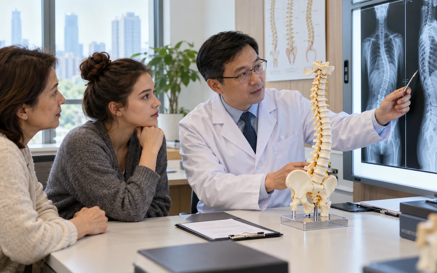
What scoliosis correction is designed to do
Scoliosis is a three-dimensional deformity: the spine curves sideways, vertebrae rotate and the body may lose balance when viewed from the side or front. Correction surgery aims to improve alignment, prevent important progression and address symptoms or functional problems while keeping the reconstruction as safe and proportionate as possible.
The common operation uses screws and rods to bring the spine toward a safer, more balanced position, followed by bone graft so the selected vertebrae fuse. The goal is not a perfectly straight X-ray. Surgeons balance correction against spinal-cord safety, lung function, bone quality and the need to preserve unfused motion.
Age, remaining growth, curve size and progression, symptoms, lung function and response to non-surgical care all matter. Adult degenerative scoliosis and adolescent idiopathic scoliosis require different decisions.
Who may be considered?
A deformity specialist may discuss surgery when the expected benefit outweighs the substantial recovery and complication risk. Situations may include:.
- A severe curve that is progressing or likely to progress substantially.
- Loss of trunk balance, pain or nerve compression that meaningfully limits daily life.
- Selected young patients whose curve cannot be adequately controlled during growth.
- Adult deformity with disabling symptoms after appropriate non-surgical treatment.
- Medical and nutritional fitness adequate for a long operation and rehabilitation.
What the hospital needs to assess
Standing full-length spine X-rays are essential because lying scans do not show balance under body weight. Bending films may show flexibility. MRI is often used when neurological symptoms, unusual curve features or spinal-cord concerns are present; CT may help with complex bone anatomy or previous fusion. The team also reviews lung function for large thoracic curves, bone density, nutrition and infection risk.
Key points for this treatment
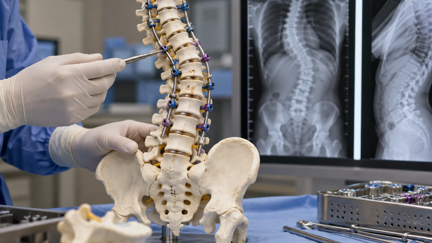
How deformity correction is performed
Most operations are performed from the back. Pedicle screws or other anchors are placed at selected levels, rods are contoured and controlled manoeuvres gradually correct rotation and balance. Bone graft is added across the intended fusion levels. Some complex curves require decompression, osteotomy or an additional approach, but these are not routine for everyone.
Neurophysiological monitoring is commonly used during surgery to watch signals from the spinal cord and nerves. The surgeon may accept less correction if monitoring, anatomy or tissue tension indicates that a more aggressive change would be unsafe.
Hospital stay and recovery
Hospital care focuses on pain control, breathing exercises, neurological checks, wound care and early safe movement. Walking usually begins with staff assistance. The return to school, work, driving and sport is staged according to the reconstruction and surgeon instructions.
Bone fusion develops over months. International patients need a clear plan for wound review, X-rays and urgent access after returning home; flight timing should be agreed with the treating team rather than based on a generic schedule.

Risks and realistic expectations
Important risks include infection, bleeding and transfusion, blood clots, lung or medical complications, spinal-fluid leak, implant problems, failure of fusion, loss of correction, persistent pain and later adjacent-level degeneration. Nerve or spinal-cord injury is uncommon but potentially serious. The risk profile is higher for very rigid curves, extensive adult reconstructions, poor bone quality and revision surgery.
New weakness or numbness, loss of bladder or bowel control, fever, wound drainage, chest pain, breathlessness or rapidly worsening pain requires urgent medical assessment.