What “minimally invasive” means
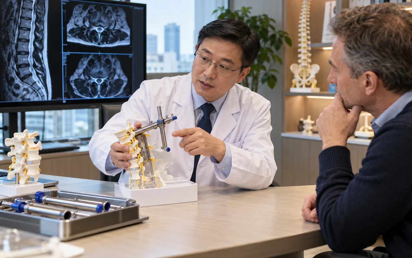
Minimally invasive spine surgery uses smaller working corridors, tubular dilators, microscopes, endoscopes or image guidance to perform selected decompressions or fusions with less disruption to surrounding muscle. The goal of surgery remains the same: treat the correctly diagnosed compression, instability or deformity.
The label covers very different operations—from a focused discectomy to instrumented lumbar fusion. It does not mean “non-surgical,” “laser” or risk-free, and a small incision can still accompany a major internal reconstruction.
Adequate decompression, safe implant placement, neurological protection and durable alignment take priority. An open approach may be safer or more complete for complex anatomy.
Who may be considered?
Smaller-access methods may be suitable for selected patients needing:.
- Microdiscectomy for a focal herniated disc.
- Laminotomy or foraminotomy for localised nerve compression.
- Selected one- or two-level lumbar fusion.
- Certain fractures, infections or deformities in experienced centres.
- Revision access where imaging and anatomy allow a safe corridor.
What the hospital needs to assess
The same complete diagnosis is required as for open surgery: symptom mapping, neurological examination, MRI, standing X-rays and CT when relevant. Body size, bone quality, prior scar, number of levels, deformity and where the compression lies determine feasibility.
Key points for this treatment
How smaller-access techniques work
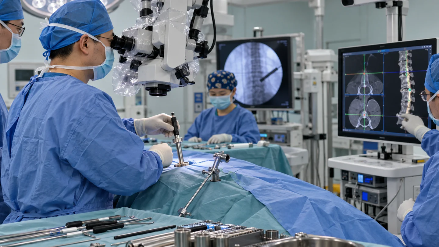
Tubular dilators separate muscle fibres to create a narrow working channel. A microscope or endoscope provides magnified vision; specialised instruments remove disc, bone or ligament. For fusion, screws may be inserted through separate small incisions with fluoroscopy or navigation, and a cage and graft are placed through the planned route.
If visibility, bleeding, anatomy or safety becomes difficult, the surgeon may enlarge the exposure or convert to an open operation. That is a safety decision, not automatically a complication.
Hospital stay and recovery
Some patients have less early muscle pain or a shorter stay, but recovery still depends on the underlying operation, neurological status and whether fusion was performed. Wound care, clot prevention, lifting restrictions and rehabilitation remain necessary.
Outcome should be judged by symptom relief, neurological function, stability and return to activity—not the number of centimetres in an incision. International patients need the same follow-up and emergency plan as after open surgery.

Risks and realistic expectations
Risks include infection, bleeding, clot, spinal-fluid leak, nerve injury, incomplete decompression, wrong-level surgery, implant malposition, non-union when fusion is performed and conversion to open surgery. Image guidance adds radiation and equipment considerations; endoscopic or tubular techniques have learning curves.
New weakness, saddle numbness, loss of bladder or bowel control, fever, wound drainage, chest pain, breathlessness or rapidly worsening pain needs urgent medical review.