
What is partial knee replacement?
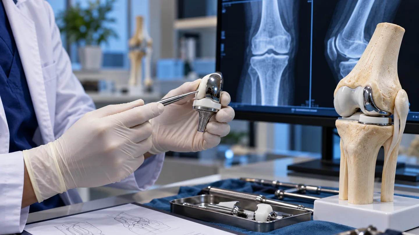
Partial knee replacement—also called unicompartmental knee arthroplasty—resurfaces only the part of the knee damaged by arthritis. The knee has medial, lateral and patellofemoral compartments. Most partial replacements treat the medial or lateral compartment between the thigh bone and shin bone; a different implant may be used when damage is confined to the kneecap compartment.
Healthy bone, cartilage and stabilising structures are preserved. This is different from total knee replacement, which resurfaces more of the joint. A smaller operation does not mean an easier selection decision: the remaining compartments, ligament function, alignment and symptoms must all support the plan.
Suitability depends on where the arthritis is located and whether the rest of the knee can continue to function. The treating surgeon must confirm that the pain pattern, examination and standing images agree.
Who may be considered?
A specialist may discuss partial replacement when pain and functional loss come predominantly from one knee compartment and non-surgical treatment no longer provides enough relief. The review commonly considers:.
- Weight-bearing X-rays showing advanced arthritis limited mainly to one compartment.
- Pain that matches the affected area rather than widespread or poorly localised symptoms.
- Usable range of motion and deformity that can be corrected during assessment.
- Ligaments that can provide the stability required by the proposed implant.
- No active infection and adequate health for anaesthesia, rehabilitation and clot prevention.
When may total replacement be more appropriate?
Widespread arthritis, major stiffness, uncorrectable deformity, inflammatory joint disease in several compartments or ligament problems may make total replacement more appropriate. Previous surgery does not automatically exclude partial replacement, but the operative history and current anatomy need careful review.
Images used for assessment
Recent weight-bearing front, side and patellar views help show which compartments are affected. A flexed weight-bearing view or full-length standing leg image may add information about joint-space loss and alignment. MRI or CT is useful in selected cases, but standing X-rays and clinical examination remain central.
Key points for this treatment
How the operation works
Under anaesthesia, the surgeon reaches the affected compartment through an incision at the front of the knee. A thin layer of damaged bone and cartilage is removed from the corresponding surfaces of the femur and tibia. Metal components cap those surfaces and a plastic bearing separates them. The surgeon then checks alignment, stability and movement before closing the wound.
What if wider damage is found?
The pre-operative discussion should cover the possibility of changing to a total knee replacement if direct inspection shows that arthritis or ligament damage is more extensive than expected. This decision should be based on the consent agreed before surgery and the findings at operation.
Conventional or robotic-assisted?
Partial replacement can be performed with conventional instruments or robotic/navigation assistance. Technology may help the team execute a planned bone cut and component position, but it does not determine whether partial replacement is appropriate. AAOS guidance reports no significant short-term difference in function, outcomes or complications between robotic-assisted and conventional knee arthroplasty, and long-term benefit remains unproven.
Hospital stay and recovery
Patients are usually encouraged to stand and walk with support soon after surgery. Pain control, wound care, clot prevention and physiotherapy begin in hospital. Some people progress faster after partial than total replacement, but discharge and recovery depend on health, balance, home support, surgical findings and the hospital’s protocol.
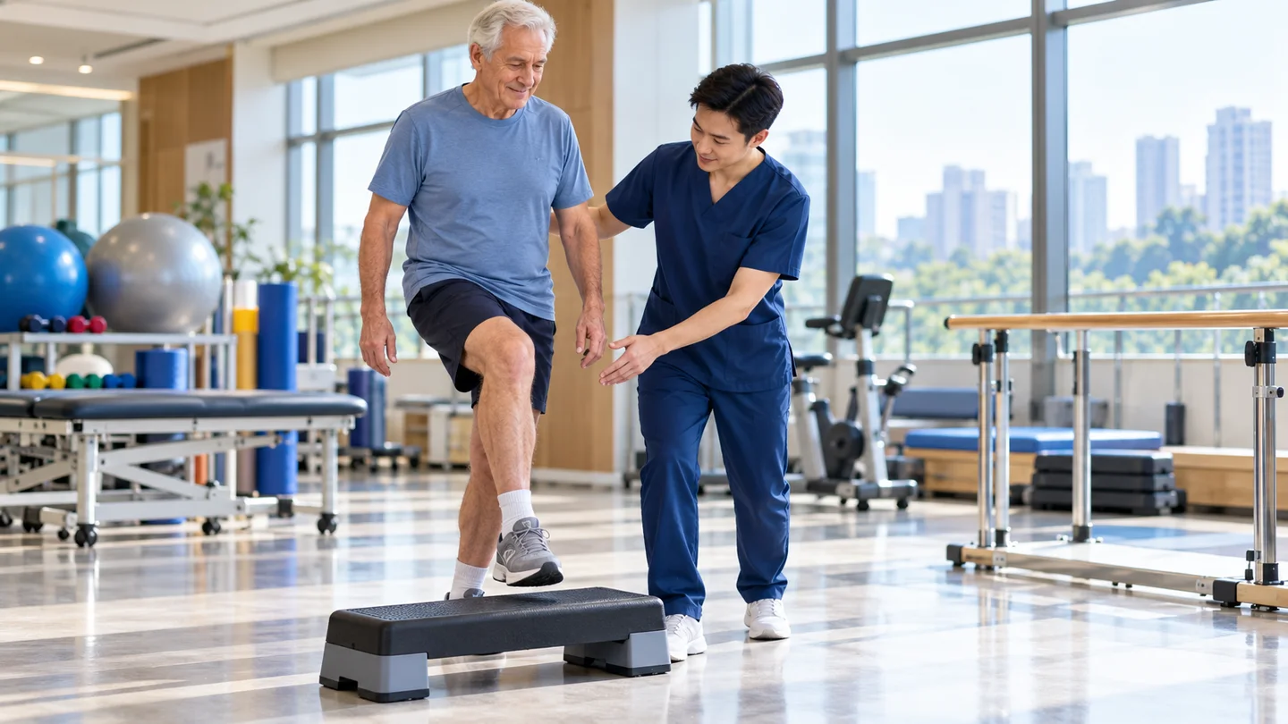
These are phases, not promised dates. Driving, work, exercise and long-haul travel require individual clearance. International patients should agree who will review the wound, prescribe medication and provide physiotherapy after returning home.
Risks and later considerations
Important risks include infection, blood clots, bleeding, anaesthetic complications, stiffness, persistent pain, nerve or blood-vessel injury, fracture, instability, bearing problems and implant loosening or wear.
Arthritis may later progress in another compartment. Some patients eventually require conversion to total knee replacement. A future operation can be more complex than a first replacement, so the surgeon should explain implant choice, expected follow-up and which symptoms need reassessment.
Sudden chest pain, shortness of breath, increasing calf swelling, high fever, wound drainage or rapidly worsening pain requires urgent clinical assessment.