Home / Neurosurgery / Skull Base Tumor Surgery
Neurosurgery patient guide · 颅底肿瘤手术

Skull Base Tumor Surgery in China

Considering skull base tumor surgery in China? Start with tumour location, prior care and specialist coordination. This guide helps you identify the relevant records, questions for the receiving team and the scope of an individual estimate before a visit is agreed.

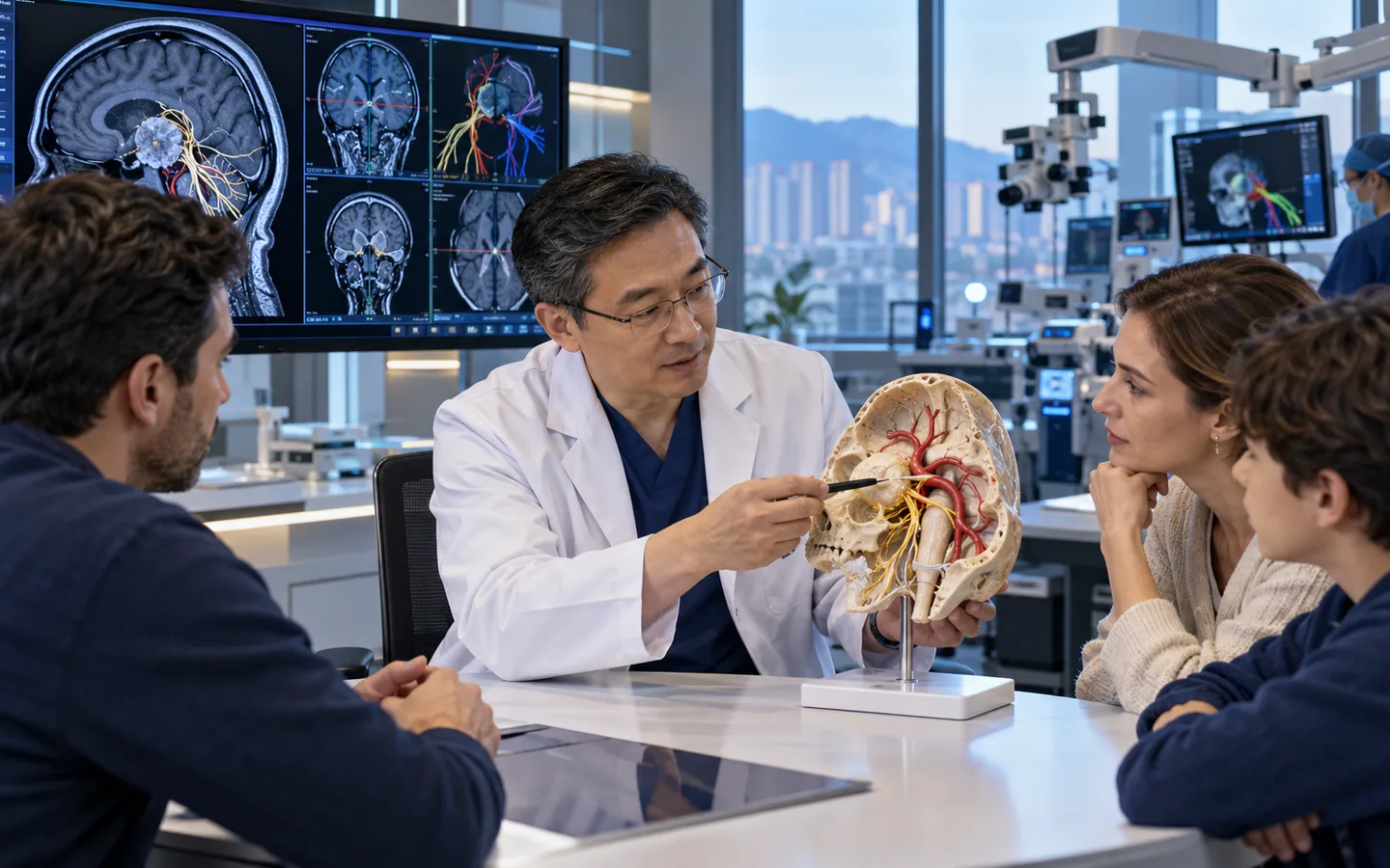
Chinese skull base neurosurgeon explaining cranial nerve and vessel anatomy to an international family

Medical records & cost enquiry

Skull Base Tumor Surgery: assessment and cost questions

For Skull Base Tumor Surgery, the budget depends on the proposed care and the hospital. A useful estimate needs to distinguish:

  • Tumour site and proposed surgical route
  • Reconstruction and specialist-team needs
  • Admission and neurological follow-up

Hospital medical fees, travel and our coordination services are separate. Any paid specialist review or coordination service is explained and agreed before you proceed.

Your next step

Start with your question

Tell us your diagnosis and what you need. Our free initial review checks the information and helps identify a suitable next step; it is not a specialist opinion or a hospital quotation.

Request a case-based estimate

Not ready to send records? Ask us first. Where hospital review is appropriate, we can help request an estimate. No travel commitment or mandatory proxy consultation.

Planning skull base tumor surgery in ChinaHospital review · individual costs · visit and follow-up

Plan the visit around tumour location, prior care and specialist coordination. Agree the assessment route before travel.

Records for the skull base tumor surgery review

Tell us what you already have: Contrast MRI brain and skull base; Thin-slice bone CT; CTA MRA or angiography if relevant. Start with a short summary; after first contact we explain which records the receiving team needs and how to share them.

Confirm the proposed scope and costs

Before asking for a personal estimate, clarify: Tumour site and proposed surgical route; Reconstruction and specialist-team needs; Admission and neurological follow-up. The receiving team confirms the proposed scope and hospital charges; coordination is agreed separately.

Visits and care after returning home

Ask which surgical and other specialists should review the case and how function-related goals and follow-up would be coordinated. Tell us if you need interpretation or English-language documents, and confirm the relevant arrangements with the receiving team.

Skull-base tumors require route-specific multidisciplinary planning

The skull base contains crowded passages for major vessels and cranial nerves controlling vision, hearing, swallowing, facial movement and other functions. Tumors include chordoma, schwannoma, meningioma and many other types.

The safest route may be endoscopic through the nose, open cranial, lateral skull base or combined. Sometimes biopsy, focused radiation or observation offers a better function-preserving strategy.

Tumor type changes the operation

A presumed diagnosis from location is not enough when tissue type would alter the route, extent or need for radiation.

Who may be considered?

Specialist review may help when the diagnosis, symptoms and previous treatment create a focused question about skull base tumor surgery.

  • A symptomatic or growing skull-base mass.
  • Cranial-nerve compression affecting vision, hearing, swallowing or facial function.
  • A tumor requiring tissue diagnosis or decompression.
  • Residual or recurrent disease after surgery or radiation.

What the specialist team must confirm

Review includes thin-slice contrast MRI and CT, vessel imaging, cranial-nerve examination, vision hearing and swallowing tests, endocrine review when relevant, systemic cancer history, pathology and prior surgery or radiation.

Key points for this treatment
Regiondeep interface of brain face and neck
Teamneurosurgery, ENT and other specialists
Planningnerve, vessel and bone anatomy
Optionssurgery, radiation or combined care
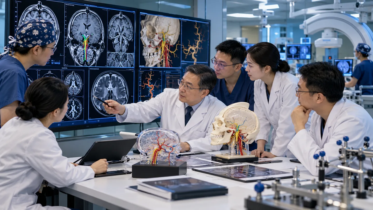
Chinese multidisciplinary skull base team planning endoscopic and open surgical corridors with MRI CT navigation
The corridor is part of the treatment decisionAccess, reconstruction and planned residual tumor are chosen to preserve critical nerves and vessels.

From anatomical mapping to a function-preserving route

The team balances tumor control against the cost of reaching and separating the lesion from critical structures.

IdentifyClarify likely biology and growth
MapDefine nerve vessel and sinus relationships
ChooseSelect route extent and reconstruction
CoordinatePlan pathology radiation and rehabilitation

Recovery is tailored to the cranial nerves at risk

Monitoring may include vision, eye movement, facial strength, hearing, swallowing, speech, shoulder function and cerebrospinal-fluid leakage.

Pathology and postoperative imaging guide surveillance or radiation. Rehabilitation may involve ENT, ophthalmology, audiology and speech or swallowing therapy.
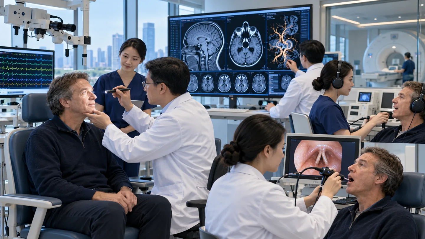
International patient completing cranial nerve hearing swallowing and MRI follow-up after skull base surgery
Function-specific follow-up detects subtle changeBaseline testing makes it possible to measure recovery and intervene early when a nerve deficit appears.
Complete resectionFollow pathology-specific surveillance
Planned residualProtect function and consider radiation
Biopsy pathwayUse diagnosis to select treatment
RehabilitationTarget cranial-nerve deficits early

Risks, limits and realistic expectations

Risks include bleeding, stroke, cranial-nerve injury, hearing or vision loss, swallowing difficulty, cerebrospinal-fluid leak, meningitis, endocrine dysfunction, incomplete removal, recurrence and death.

Do not delay urgent local care

New vision loss, weakness, choking, severe headache, clear nasal or ear fluid, fever or reduced consciousness needs urgent local care.

Before hospital review

Records for skull base tumor surgery assessment

A safe international review depends on dated source reports, original imaging and a complete treatment timeline—not a diagnosis label alone.

Contrast MRI brain and skull base
Thin-slice bone CT
CTA MRA or angiography if relevant
Vision hearing and cranial-nerve tests
Swallowing and speech assessment
Original pathology slides or blocks
Prior operation and radiation records
Endocrine testing when relevant

Tell us what you need

Ask about your care,
your hospital and your budget.

You can ask about suitability, an expert opinion, an appointment or the likely medical cost. If you are unsure, choose “Not sure — please advise”.

This enquiry is aboutSkull Base Tumor SurgeryNot sure — please advise

How a personal estimate is prepared

  1. Tell us about your case.Describe your diagnosis, main question and preferred city, if any.
  2. Share the relevant records.We explain what is needed and how to send it by WhatsApp or email.
  3. Request a hospital estimate.Where appropriate, we help request hospital review and a cost estimate. Any paid review is agreed first.

This is an enquiry, not an order or payment. Proxy consultation is not mandatory. Any service scope is agreed separately before you proceed.

Ask about Skull Base Tumor Surgery

A first enquiry is free. Email and permission to respond are required; the other details are optional. Any paid clinical review or coordination is discussed separately.

Included automatically so we know which procedure you are asking about.
A preference, not a confirmed appointment.
Please do not send passport numbers, card details or full medical files in this first enquiry. We will explain which records are needed next.

Send a short summary first. This is an enquiry, not an order, payment or confirmed appointment.

No booking or payment is made by sending this enquiry.
Editorial transparency

Medical sources

Patient information is based on established government and professional guidance. Content updated 5 October 2026. This is patient information, not an individual clinical assessment.